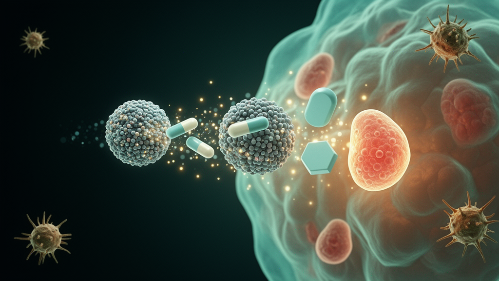
Researchers have built a reactive oxygen species-responsive nanoparticle that shuttles a PARP inhibitor prodrug and a DOT1L histone methyltransferase inhibitor into ovarian cancer cells at the same time, and the combination substantially outperformed the PARP inhibitor alone in cell and mouse models. The platform, described in Materials Today Bio, is designed to break through two known limits of PARP inhibitor therapy: restored DNA repair capacity and active pumping of the drug back out of tumor cells.
The rationale rests on a signaling chain that the authors summarize as the PARP1/DOT1L/H3K79me2-PLCG2/ABCB1 axis. DOT1L is the only known histone methyltransferase that methylates lysine 79 on histone H3, a modification tied to DNA repair and transcriptional regulation, and it is highly expressed in many malignancies. In ovarian cancer, public transcriptomic datasets analyzed by the team showed significant DOT1L upregulation in tumor tissue compared with normal tissue, and immunohistochemical data from the Human Protein Atlas showed elevated staining intensity in tumors. Kaplan-Meier analysis indicated that elevated DOT1L levels were associated with poorer clinical outcomes, and drug response profiling revealed a positive correlation between sensitivity to the DOT1L inhibitor SGC0946 and sensitivity to PARP inhibitors.
Mechanistically, the team found that DOT1L protein was significantly upregulated in PARP inhibitor-resistant A2780 cells compared with parental lines, and that co-treatment with the DOT1L inhibitor reduced PARP inhibitor efflux and boosted intracellular drug accumulation in resistant cells, measured by high-performance liquid chromatography. In four ovarian cancer cell lines (A2780, ID8, SK-OV-3 and OVCAR-8), the combination of DOT1L inhibitor and PARP inhibitor produced significantly greater killing than either drug alone, and in A2780 cells the combination drove an apoptosis rate of 29.88 percent, versus 10.99 percent for the DOT1L inhibitor and 14.56 percent for the PARP inhibitor alone.
Building on that synergy, the team engineered the delivery system. A Niraparib prodrug was synthesized by conjugating the clinically approved PARP inhibitor to a ROS-responsive monomer through a thioacetal linkage, and a phenylboronic acid-functionalized polymer was designed to load SGC0946. The two polymers were co-assembled at a 1:1 ratio into a nanoparticle, with 10 percent by weight of the DOT1L inhibitor, yielding the final construct, DP@NP. Transmission electron microscopy showed uniform spherical particles with average diameters of roughly 96.8 nanometers for DP@NP and 123.2 nanometers for the drug-free assembly, while dynamic light scattering gave hydrodynamic diameters of 116.4 and 131.4 nanometers respectively, with polydispersity indices of 0.112 and 0.061 indicating tight size uniformity.
The ROS trigger is central to the design. When exposed to hydrogen peroxide at 10 millimolar, a concentration used to mimic the oxidative tumor microenvironment, the thioacetal linkage cleaves and a self-immolative reaction releases the parent drug Niraparib. HPLC analysis showed that the prodrug was almost entirely converted to Niraparib after 48 hours at 37 degrees Celsius, and mass spectrometry confirmed the appearance of the Niraparib ion peak. Under the same oxidative conditions, DP@NP released about 94.9 percent of its PARP inhibitor payload within 48 hours, whereas release in phosphate-buffered saline remained minimal at 25.6 percent. The nanoparticles also disassembled almost completely in hydrogen peroxide, with TEM showing the spherical structures nearly absent after exposure, and they remained size-stable in buffer over seven days.
In cell studies, DP@NP was internalized by A2780 cells in a time-dependent manner, with mean fluorescence intensity at 7 hours roughly 1.5-fold and 3.8-fold higher than at 4 and 1 hours, and confocal imaging showed the label accumulating mainly in the cytoplasm. The cytotoxicity gains were striking: the half-maximal inhibitory concentration of DP@NP in A2780 cells was 1.32 micromolar, compared with 14.3 micromolar for free PARP inhibitor, and the mean apoptosis rate reached 53.29 percent for DP@NP versus 22.72 percent for the drug-free nanoparticle and 15.0 percent for free drug. DP@NP also penetrated and killed cells throughout three-dimensional tumor spheroids and showed the strongest inhibition of colony formation among all groups.
The mechanistic assays traced these effects to the intended targets. Western blotting showed that DP@NP markedly elevated the DNA damage marker gamma-H2AX while downregulating DOT1L and the drug efflux transporter ABCB1. Immunofluorescence revealed weaker ABCB1 signal in DP@NP-treated cells, and HPLC quantification showed intracellular PARP inhibitor levels 2.6-fold and 4.6-fold higher than with the drug-free nanoparticle and free drug respectively. Gamma-H2AX levels rose about 1.4-fold and 1.2-fold above the free drug and nanoparticle groups, and cytosolic double-stranded DNA accumulated visibly, consistent with impaired DNA repair and reduced drug efflux converging to amplify damage.
That damage translated into innate immune signaling. DP@NP produced the strongest p-STING fluorescence among the treatments, roughly 1.5-fold and 1.2-fold above free drug and drug-free nanoparticle, and increased p-TBK1 and p-IRF3, the downstream arms of the STING-TBK1-IRF3 pathway. ELISA showed a 2.8-fold increase in IFN-beta and a 1.7-fold increase in TNF-alpha compared with free PARP inhibitor. When treated ID8 cells were co-cultured with bone marrow-derived dendritic cells from female C57BL/6 mice, DP@NP drove a mature dendritic cell proportion of 43.5 percent, marked by upregulated CD80 and CD86. The treatment also raised PD-L1 expression on tumor cells, a known consequence of PARP inhibitor activity that can be exploited with checkpoint blockade. RNA sequencing of treated A2780 cells confirmed significant DOT1L knockdown and enrichment of DNA damage response, p53 signaling, interferon-related signaling and IL6-JAK-STAT3 pathways.
In vivo, the platform was tested in a high-grade serous ovarian cancer patient-derived xenograft model, which retains the histopathological and genetic features of the primary tumor. After tail vein injection of fluorescently labeled nanoparticles, tumor signals appeared within 4 hours, peaked around 24 hours, and remained strong at 48 hours, with accumulation primarily in tumors, liver and kidneys. Mice received intravenous injections on days 1, 3, 5, 7 and 9. By day 13, DP@NP achieved an 87.3 percent tumor inhibition rate, compared with 61.3 percent for the drug-free nanoparticle and 38.0 percent for free PARP inhibitor, and the mean tumor weight in the DP@NP group was 0.12 grams versus 0.64 grams for free drug and 0.41 grams for the nanoparticle without the DOT1L inhibitor. Body weight stayed stable across groups, serum biochemistry showed no significant differences, and organ histology revealed no abnormalities, supporting favorable tolerability in these models.
In an ID8-Luc peritoneal metastasis model of advanced ovarian cancer, bioluminescence in the control group by day 16 was approximately 11.0 times higher than in the DP@NP group, and immune profiling of ascites showed mature dendritic cells at 53.0 percent versus 22.7 percent with free drug, reduced myeloid-derived suppressor cells at 34.8 percent versus 51.6 percent with the drug-free nanoparticle, elevated NK cells at 71.1 percent, and a shift of macrophages toward the M1 phenotype. PD-1-positive CD8+ T cells and PD-L1 both rose, suggesting immune activation accompanied by checkpoint engagement. Combining DP@NP with an anti-PD-L1 antibody produced the lowest tumor burden in the ascites model, raised splenic central memory T cells to 28.4 percent, and increased CD8+ T cell infiltration in ascites to 48.9 percent. The authors caution that long-term safety characterization, scalable manufacturing, variability in tumor ROS levels between patients, and validation in more clinically relevant models remain challenges before clinical translation.
Subject of Research: A ROS-responsive nanoparticle co-delivering a PARP inhibitor prodrug and a DOT1L inhibitor to amplify cGAS-STING-driven antitumor immunity in ovarian cancer.
Article Title: Targeting histone methylation with ROS nanoparticles delivering PARPi prodrugs to amplify the stimulation of cGAS-STING for immunotherapy in ovarian cancer
Article References: Sun, Y., Li, H., Zhang, H., Zhang, H., Xu, C., Li, B., Xiao, H., & Zhao, D. (2026). Targeting histone methylation with ROS nanoparticles delivering PARPi prodrugs to amplify the stimulation of cGAS-STING for immunotherapy in ovarian cancer. Materials Today Bio, 41, Article 103777. https://doi.org/10.1016/j.mtbio.2026.103777
Image Credits: AI Generated
DOI: 10.1016/j.mtbio.2026.103777
Keywords: ovarian cancer, PARP inhibitors, DOT1L, cGAS-STING, nanomedicine, ROS-responsive nanoparticles, immunotherapy, anti-PD-L1, drug efflux, DNA damage, tumor microenvironment, preclinical study
News Source: Nathaniel Bowman. (October 28, 2026). ROS-responsive nanoparticle pairs PARP inhibitor with DOT1L blocker to fire up antitumor immunity in ovarian cancer models. Scienmag.