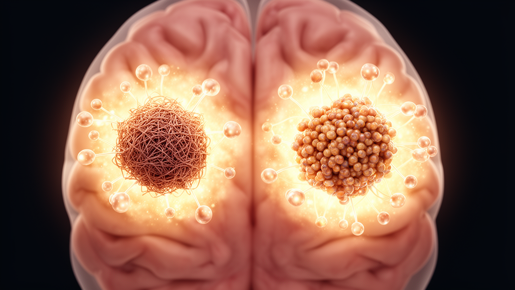
Amyloid plaques, one of the defining neuropathological features of Alzheimer’s disease, are built from insoluble amyloid beta (Aβ) peptides. But not all Aβ is the same. The peptide undergoes post-translational modifications that change how it aggregates, how resistant it is to breakdown, and how toxic it becomes, producing a heterogeneous mixture of species that may contribute differently to the disease process. One of the most consequential variants is pyroglutamate-modified Aβ at glutamate position 3, known as pE3-Aβ, an aggregation-prone and proteolytically resistant form that preferentially accumulates within plaque cores, has been implicated in early plaque seeding, and is a major target of emerging anti-amyloid immunotherapies.
Despite that importance, the molecular environment surrounding pE3-Aβ, compared with the broader pool of Aβ species detected by the widely used 6E10 antibody, has remained incompletely defined in human tissue.
The researchers used a technique called Biotinylation by Antibody Recognition, or BAR, an in-situ proximity labeling approach. The method works in formalin-fixed postmortem tissue: an antibody recognizes its target within the preserved section, and a labeling reaction tags proteins in the immediate molecular vicinity.
The team applied BAR to hippocampal tissue from pathologically confirmed Alzheimer’s disease cases and from cognitively normal controls, probing for pE3-Aβ and for 6E10-positive amyloid pathology in each group. Differential proteomic analysis of the resulting captures identified 48 significantly enriched proteins in AD pE3-Aβ captures, 28 in AD 6E10 captures, and 15 in 6E10 captures from cognitively normal tissue. Strikingly, no significant enrichment was detected in pE3-Aβ captures from the control group, a result the authors say supports the view of pE3-Aβ as a pathology-associated species rather than a feature of normal aging tissue.
The comparison also revealed how much of each signature was unique. pE3-Aβ in Alzheimer’s disease showed the largest variant-specific profile, with 31 proteins found only in its captures. The 6E10-positive amyloid had 11 unique proteins in AD tissue and 14 unique proteins in cognitively normal tissue. Sixteen proteins were shared between AD pE3-Aβ and AD 6E10 captures, and one protein, PCSK1N, was shared across AD pE3-Aβ and both the AD and control 6E10 captures, marking it as a common neighbor of amyloid regardless of disease state or antibody target.
Beyond individual proteins, pathway enrichment analysis of the proximal proteomes pointed to broader biological disruptions linked to pE3-Aβ. These included synaptogenesis signaling, clathrin-mediated endocytosis, mitochondrial division signaling, and neurotransmitter release. In other words, the proteins clustering around this modified peptide were disproportionately involved in how synapses form, how cells internalize material through coated vesicles, how mitochondria divide, and how chemical signals are released between neurons.
Some pathways were shared across amyloid forms rather than specific to one variant. These convergent pathways suggest that multiple species of plaque-associated Aβ sit near overlapping synaptic and signaling machinery in the diseased hippocampus.
The study also took advantage of the control tissue to ask whether ordinary, age-associated amyloid deposition carries a different protein entourage than pathological deposition.
The authors frame their findings as evidence that pE3-Aβ is associated with an expanded, variant-specific proximal proteome in human AD hippocampus, and as a possible indication that intracellular trafficking, synaptic signaling, and mitochondrial pathways represent network-level vulnerabilities relevant to pE3-Aβ pathology. Because the work was performed in postmortem, formalin-fixed tissue, it captures spatial associations rather than demonstrating causal effects, and the proximity labeling reflects molecular neighborhoods at the time of death. Even so, by working directly in human hippocampal tissue and separating a chemically distinct Aβ variant from the general amyloid pool, the approach offers a template for dissecting which molecular environments accompany which forms of amyloid as Alzheimer’s disease takes hold.
Subject of Research: Mapping the proximal proteomes of pE3-Aβ and 6E10 amyloid pathology in human Alzheimer's disease hippocampus
Article Title: Proximity labeling reveals unique and shared proximal proteomes associated with pE3-Aβ and 6E10 amyloid pathology in human Alzheimer’s disease hippocampus
Article References: Alia, A., Urquhart, K., Carson, H., Killinger, B., Janson, C., & Romanova, L. (2026). Proximity labeling reveals unique and shared proximal proteomes associated with pE3-Aβ and 6E10 amyloid pathology in human Alzheimer’s disease hippocampus. Acta Neuropathologica Communications. https://doi.org/10.1186/s40478-026-02441-5
Image Credits: AI Generated
DOI: 10.1186/s40478-026-02441-5
Keywords: Alzheimer's disease, amyloid beta, pE3-Aβ, proximity labeling, hippocampus, proteomics, BAR, postmortem tissue, synaptic signaling, mitochondrial pathways, plaque cores, neurodegeneration
News Source: Diana Fleming. (October 30, 2026). Proximity Labeling Maps the Protein Neighborhoods of Two Amyloid Forms in Alzheimer’s Hippocampus. Scienmag.