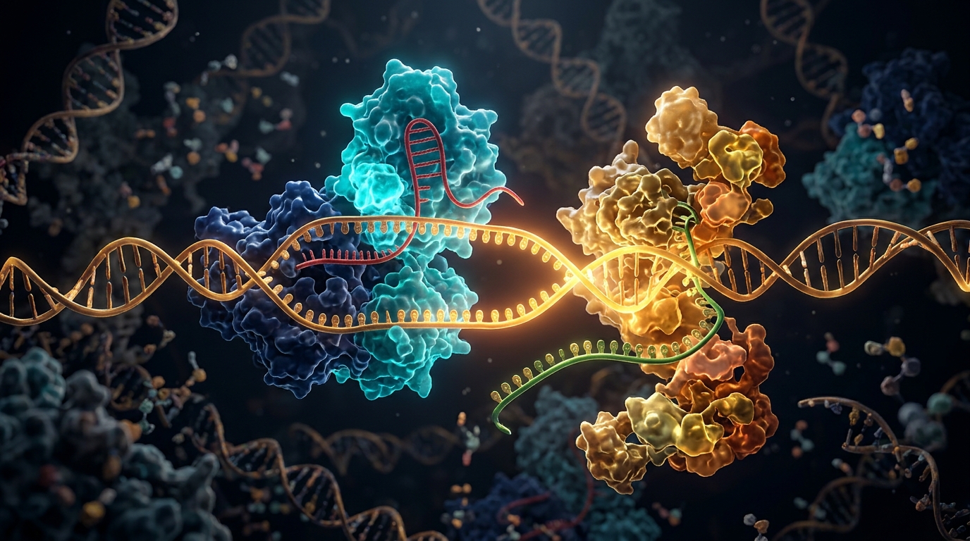
Scientists have captured detailed three-dimensional snapshots of a molecular machine that can write new genetic cargo into a chosen spot in the human genome, revealing for the first time how the pieces of the system fit together as the insertion reaction begins. In a study published in Nature Structural & Molecular Biology, a team led by Daisuke Komatsu and Hiroshi Nishimasu of The University of Tokyo, together with Jonathan Gootenberg and Omar Abudayyeh of the Massachusetts Institute of Technology, used cryo-electron microscopy to determine the structures of the R2 retrotransposon from the zebra finch Taeniopygia guttata, both on its own and attached to a Cas9–guide RNA complex. The work provides a mechanistic framework for STITCHR, a programmable gene-integration system that couples the retrotransposon to CRISPR–Cas9, and the researchers used those structural insights, combined with computational protein design, to engineer an enhanced version of the editor.
R2 belongs to a class of non-long-terminal-repeat retroelements that insert themselves almost exclusively into 28S ribosomal DNA loci across a wide range of eukaryotes. These elements home in on their targets through a process called target-primed reverse transcription, or TPRT. In TPRT, the R2 protein recognizes and nicks one strand of the target DNA to create a primer, then uses its own RNA as a template to synthesize DNA directly onto that primer. The avian R2 system from Taeniopygia guttata, abbreviated R2Tg, is notable because it can insert a payload sequence carried within its RNA template into 28S ribosomal DNA loci even in human cells, a property that made it an attractive starting point for engineering.
Before solving the structures, the team characterized the biochemistry of R2Tg in vitro. They mixed purified R2Tg protein with its cognate RNA template and a fluorescently labeled 280-base-pair 28S ribosomal DNA target, with or without nucleotide building blocks, and analyzed the products on denaturing gels. R2Tg cleaved the bottom strand of the target DNA and catalyzed the TPRT reaction in the presence of deoxynucleotides, producing two major products longer than 500 nucleotides; the larger products arise from template jumping, in which DNA synthesis continues using another RNA molecule after the completion of TPRT. Compared with the classical silkworm retrotransposon R2Bm, R2Tg produced both larger template-jumping products and smaller TPRT products more efficiently, suggesting that its higher TPRT activity drives more template jumping. Intriguingly, while R2Bm primarily cleaved the bottom strand, R2Tg cleaved both the top and bottom strands, highlighting a mechanistic difference between the two retrotransposons. The experiments also defined the RNA requirements: R2Tg needed the 3′ untranslated region of its template but neither the 5′ untranslated region nor the 5′ homology arm, required at least a 4-nucleotide 3′ homology arm for efficient TPRT, and depended on a roughly 100-nucleotide stretch of the 3′ untranslated region upstream of that homology arm.
To visualize the reaction, the researchers reconstituted a ternary complex containing R2Tg, a 115-nucleotide template RNA and an 80-base-pair target 28S ribosomal DNA, adding a chain-terminating nucleotide, ddTTP, that would be incorporated into the nicked bottom strand as the first nucleotide to stall DNA synthesis at a defined point. They then determined cryo-EM structures of the complex in three distinct functional states at resolutions between 2.2 and 2.6 angstroms. Because the first state was the predominant one and most protein domains were resolved at 2.2-angstrom resolution, the team focused their description on that state. The structures revealed that the R2Tg protein is organized into an N-terminal DNA-binding region containing three zinc-finger domains and a Myb domain, connected by a spacer region to a reverse-transcriptase–restriction-enzyme-like enzymatic core that includes an N-terminal extension domain, an RT domain, a thumb domain, a linker domain, a zinc-knuckle domain and an RLE domain. The RT and thumb domains form a right-hand-like architecture that cradles a template RNA–primer DNA heteroduplex, and densities for the incorporated ddTTP and an incoming nucleotide in the RT active site confirmed that the captured structure represents the TPRT initiation state.
One of the most striking findings concerns how R2Tg reads its RNA template. Only the 3′ 61 nucleotides of the 3′ untranslated region, together with the adjacent 3′ homology arm, are visible in the structure, and this core adopts a highly specific architecture: a pseudoknot with two stems, a stem loop, and two single-stranded linker regions. The pseudoknot is stabilized by three successive base triples and is sandwiched between the ZF3 and NTE−1 domains of the protein. Functional tests reinforced the structural picture: a 66-nucleotide 3′ untranslated region supported TPRT in vitro just as well as the full-length 325-nucleotide version, whereas an additional 10-nucleotide deletion abolished reverse transcription without affecting DNA cleavage, and an RNA mutant in which the unrecognized upper portion of the stem loop was replaced with a GAAA tetraloop still supported TPRT. Together, these data demonstrate that the RNA 3′ untranslated region core adopts a unique structure recognized by R2Tg, which positions the 3′ homology arm to base-pair with the nicked bottom strand and prime reverse transcription.
The structures also explain how the protein finds and opens its DNA target. The target DNA remains double-stranded upstream and downstream of the cleavage site but is unwound and split into two separate strands around the bottom-strand nick. Multiple domains make base-specific contacts with the 28S ribosomal DNA: residues in the ZF2, RT, Myb and ZF–Myb linker regions hydrogen-bond with particular bases, consistent with R2Tg’s known preference for the 28S target. Meanwhile, the zinc-kuckle and RLE domains form a wedge-like protrusion that inserts into the DNA, with two residues stacking against a base pair to promote unwinding around the cleavage site. Mutational tests showed that individual disruptions at three protein-contacting sites in the target DNA did not impair TPRT, but simultaneous mutation of all three abolished activity, and an R2Tg mutant lacking its DNA-binding domains showed no TPRT at all. A mutation around the cleavage sites eliminated top-strand cleavage while sparing bottom-strand cleavage, indicating that base-specific recognition by the RLE active site matters for the second-strand cut but not the first.
With the natural system mapped, the team turned to the engineered STITCHR system, in which a Cas9 nickase cuts one strand of a chosen genomic site and R2Tg then uses that cut strand as the primer for reverse transcription of a separate template RNA. The researchers reconstituted a five-component complex containing a catalytically inactive Cas9, a guide RNA, a prenicked non-28S target DNA, R2Tg and a template RNA, and determined its cryo-EM structure at 3.3-angstrom resolution. In the structure, Cas9 grips the guide RNA and target DNA as in previously reported Cas9 structures, while the R2Tg–template RNA complex sits adjacent to it, with the R2Tg RLE domain touching the Cas9 RuvC domain. The nicked bottom strand threads through a channel between the R2Tg linker domain and the Cas9 RuvC domain, a geometry that facilitates efficient handover of the DNA primer from the Cas9 complex to the retrotransposon complex. Notably, the non-28S target DNA in this engineered setting is recognized by the ZF3/2 and ZF1 domains through base-specific contacts that arise coincidentally at key positions, supporting the idea that R2Tg can be reprogrammed to novel sites as long as the cleaved strand base-pairs sufficiently with the template RNA’s 3′ homology arm.
High-speed atomic force microscopy added a dynamic dimension to the static structures. Movies of the STITCHR complex containing a 76-nucleotide template RNA confirmed that most molecules adopt a conformation similar to the cryo-EM structure, while also revealing flexibility in the arrangement between Cas9 and R2Tg. Complexes carrying a longer, 335-nucleotide template RNA showed the upstream portion of the RNA projecting flexibly from the R2Tg molecule, consistent with the notion that the structured 3′ untranslated region core binds the protein at the initiation stage while the rest of the template remains mobile.
The structural insights then guided engineering. Comparing configurations in human cells, the team found that expressing Cas9 and R2Tg separately, rather than as a fusion, achieved roughly 1.5-fold higher integration efficiencies at reprogrammed genomic loci, so they adopted the separated system for subsequent work. Deletion experiments revealed unexpected sensitivity: removing the ZF3/2 and ZF1 domains abolished STITCHR-mediated integration, while deleting the Myb domain reduced it, pointing to functional importance of the interactions between these domains and the enzymatic core. Using protein language models, the researchers identified point mutations such as N640A, E1027K and E1027R that enhanced both R2Tg and STITCHR activities, and ProteinMPNN-based design of the Cas9–R2Tg interface yielded mutants, notably M2, that improved integration efficiencies at both the AAVS1 and NOLC1 sites by approximately 1.5 to 2-fold over the wild-type editor. Combining the best interface mutations with E1027R produced the top-performing variant, named e-STITCHR, which enables reprogrammed integration with approximately 2 to 2.5-fold higher efficiency relative to the original system.
The study also situates STITCHR among genome-editing technologies. A structural comparison with prime editing, which couples Cas9 nicking to reverse transcription by an engineered viral reverse transcriptase tethered to an extended guide RNA, highlights a key architectural difference: in prime editing the RNA template is physically linked to the guide, which makes accommodating long RNA–DNA products difficult, whereas in STITCHR the Cas9 guide RNA and the R2 template RNA are separate molecules, and a structural model suggests the R2 complex can dissociate from Cas9 after reverse transcription initiation, allowing long heteroduplex production without steric hindrance. This helps explain why STITCHR can integrate sequences longer than 10 kilobases while prime editing typically inserts less than 50 base pairs. The authors note that engineering a STITCHR system based on an R2 ortholog from Talpa occidentalis, which shows reprogrammed integration with minimal insertion at its natural 28S target in human cells, will be an important direction for future work. For now, the structures deliver what the field has lacked: an atomic-level view of how a retrotransposon primes, positions and begins writing DNA, and a blueprint for making that writing faster, more precise and more programmable.
Subject of Research: Cryo-EM structures of the R2Tg retrotransposon in its target-primed reverse transcription initiation state, alone and coupled to CRISPR–Cas9 in the STITCHR gene-integration system
Article Title: Structural mechanism of programmable gene integration by the R2 retrotransposon coupled with CRISPR–Cas9
Article References: Komatsu, D., Hiraizumi, M., Fell, C. W., Fujimatsu, K., Nagahata, N., Yamada, S., Sapkota, S., Ramani, H., Arya, B., Shibata, M., Yamashita, K., Gootenberg, J. S., Abudayyeh, O. O., & Nishimasu, H. (2026). Structural mechanism of programmable gene integration by the R2 retrotransposon coupled with CRISPR–Cas9. Nature Structural & Molecular Biology. https://doi.org/10.1038/s41594-026-01903-7
Image Credits: AI Generated
DOI: 10.1038/s41594-026-01903-7
Keywords: R2 retrotransposon, cryo-EM, target-primed reverse transcription, STITCHR, CRISPR–Cas9, gene integration, 28S ribosomal DNA, reverse transcriptase, protein engineering, genome editing, Taeniopygia guttata, prime editing
News Source: Juliet Wilcox. (November 4, 2026). Cryo-EM reveals how a retrotransposon and CRISPR–Cas9 team up to insert genes. Scienmag.