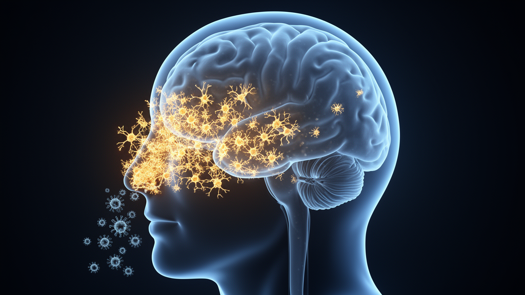
Herpes simplex virus type 1 is best known for causing cold sores, but the virus is also a neurotropic pathogen with the ability to invade the central nervous system, where it has been linked to chronic neuroinflammation, cognitive impairment, and an elevated risk of neurodegenerative disease. A central question in neuroimmunology has been how the brain’s resident immune cells, the microglia, respond to this invasion at the molecular level, and why their inflammatory activity can persist long after the initial infection. A new study published in PLOS Pathogens offers the most detailed answer yet, using an unusually comprehensive combination of single-cell, epigenetic, and spatial technologies to map the brain’s immune response to intranasal HSV-1 infection in unprecedented resolution.
The research team, led by Seth Frietze and Christy S. Niemeyer along with colleagues including Diego Restrepo and Maria A. Nagel, set out to define the transcriptional and epigenetic mechanisms that shape microglial behavior during acute HSV-1 infection in a living animal model. Rather than studying isolated cells in a dish, which can distort immune cell behavior, the investigators analyzed brains directly following intranasal infection, the natural route by which the virus can enter through the nasal cavity and reach the brain. This in vivo approach is critical because microglia are highly sensitive to their tissue environment, and their responses to infection are shaped by signals from neighboring neurons, astrocytes, and infiltrating immune cells.
The methodological core of the study is a multiomic design that, according to the authors, has never before been applied to this infection model. The team performed single-nucleus RNA sequencing alongside measurements of chromatin accessibility, a technique that reveals which regions of DNA are open and available for regulatory proteins to bind. They focused on CD11b-positive nuclei, a molecular marker that captures microglia and other myeloid immune cells of the brain. By pairing gene expression data with chromatin accessibility profiles from the same cells, the researchers could identify not only which cell populations existed in the infected brain but also which regulatory programs were being switched on within each population.
The single-cell analysis revealed a strikingly heterogeneous landscape. Rather than a uniform population of activated immune cells, the infected brains contained transcriptionally and epigenetically distinct microglial and macrophage-associated populations. HSV-1 infection redistributed these states, shifting the balance away from homeostasis and toward an overrepresentation of interferon-responsive microglia and macrophage-like cells. Interferons are the body’s first-line antiviral cytokines, and their signature was unmistakable: the responsive populations showed amplification of the transcription factors STAT1 and STAT2, IRF1, and CEBPB, a trio of regulators that together orchestrate the cellular response to type I interferon signaling.
Equally telling was what disappeared. Homeostatic microglial gene signatures, including well-established markers such as ApoE and Cst3, were reduced following infection. Homeostatic microglia perform essential housekeeping functions in the healthy brain, surveying their surroundings, pruning synapses, and supporting neuronal metabolism. A loss of these signatures suggests that infection does not simply add an inflammatory program on top of normal microglial function but instead fundamentally remodels the cells, trading their maintenance roles for antiviral and inflammatory states. This kind of trade-off has important implications, because prolonged suppression of homeostatic functions is a feature shared by several chronic neurodegenerative conditions.
One of the study’s most significant contributions is its spatial dimension. Single-cell sequencing, while powerful, typically requires dissociating tissue, which destroys information about where each cell was located. To recover that spatial context, the researchers turned to spatial transcriptomics, a method that maps gene expression directly onto intact tissue sections. Their analysis localized HSV-1 antigen to discrete regions of the brainstem, revealing that the infection was not diffusely spread throughout the brain but concentrated in anatomically defined areas. This finding aligns with known routes of intranasal viral entry, in which the virus can travel along olfactory and trigeminal nerve pathways into the central nervous system.
The spatial data did more than confirm where the virus was. The brainstem regions harboring viral antigen were precisely the regions enriched for the predicted targets of STAT1/2, IRF1, and CEBPB regulation identified in the single-nucleus analysis. In other words, the interferon-driven regulatory networks inferred from isolated nuclei were demonstrably active within the anatomical territories where the virus resided. This convergence between single-cell inference and spatial validation is a methodological strength, because it demonstrates that the regulatory programs detected at the single-cell level are not artifacts of tissue processing but reflect genuine, spatially restricted biology in the infected brain.
Complementary spatial proteomics reinforced the picture. Protein-level measurements confirmed increased microglial activation and astrocytic responses specifically within the HSV-1 antigen-positive regions. Astrocytes, the star-shaped glial cells that support neurons and help maintain the blood-brain barrier, are known to adopt reactive states during infection and injury. Their localized activation alongside activated microglia indicates that the interferon-dominant response recruits multiple glial cell types within the same infected territories, creating focal pockets of intense neuroinflammation surrounded by relatively spared tissue.
Taken together, the integrated analyses demonstrate that intranasal HSV-1 infection induces a localized, transcriptionally and epigenetically defined remodeling of microglia that is dominated by interferon-driven regulatory programs and accompanied by a loss of homeostatic identity. The emphasis on region specificity matters: the brain’s response to the virus is not a global inflammatory surge but a mosaic of focal immune activation tied to the anatomical distribution of viral antigen. This localized architecture could help explain how a transient infection produces lasting neurological consequences, since repeatedly or chronically activated microglia in discrete circuits may gradually erode neuronal health in those circuits while leaving other regions comparatively intact.
The findings carry broader implications for understanding the relationship between viral infection and neurodegenerative disease. Epidemiological and experimental studies have increasingly implicated HSV-1 in Alzheimer’s disease and other dementias, but the mechanistic bridge between acute infection and chronic pathology has remained elusive. By linking single-cell regulatory architecture to spatially restricted tissue responses, this study provides a mechanistic framework: localized interferon-dominant microglial remodeling, sustained by epigenetic changes in chromatin accessibility, may represent the cellular substrate through which viral invasion seeds long-term neurological vulnerability. The epigenetic dimension is particularly noteworthy, because chromatin remodeling can persist after the initial stimulus has resolved, potentially locking immune cells into a primed state. While the study focuses on acute infection, its integrated multiomic approach establishes a template for future work examining how these region-specific immune programs evolve over time, whether they can be reversed, and whether targeting the STAT1/2-IRF1-CEBPB axis might one day mitigate the neurological toll of herpesvirus infections in the brain.
Subject of Research: Microglial transcriptional and epigenetic responses to intranasal HSV-1 infection in the brain
Article Title: Intranasal HSV‑1 infection drives region‑specific interferon‑dominant microglial remodeling
Article References: Frietze, S., Lunn, C., Oldham, D., Boyd, J. R., Bubak, A. N., Saucedo, A. B., Nagel, M. A., Restrepo, D., Bruce, K. D., & Niemeyer, C. S. (2026). Intranasal HSV‑1 infection drives region‑specific interferon‑dominant microglial remodeling. PLOS Pathogens, 22(10), e1014684. https://doi.org/10.1371/journal.ppat.1014684
Image Credits: AI Generated
DOI: 10.1371/journal.ppat.1014684
Keywords: HSV-1, microglia, neuroinflammation, interferon, single-nucleus RNA sequencing, spatial transcriptomics, chromatin accessibility, brainstem, STAT1, neurodegenerative disease, astrocytes, PLOS Pathogens
News Source: Kristina Jarvis. (October 14, 2026). Intranasal Herpes Virus Remodels Brain Immune Cells in a Region-Specific Pattern. Scienmag.