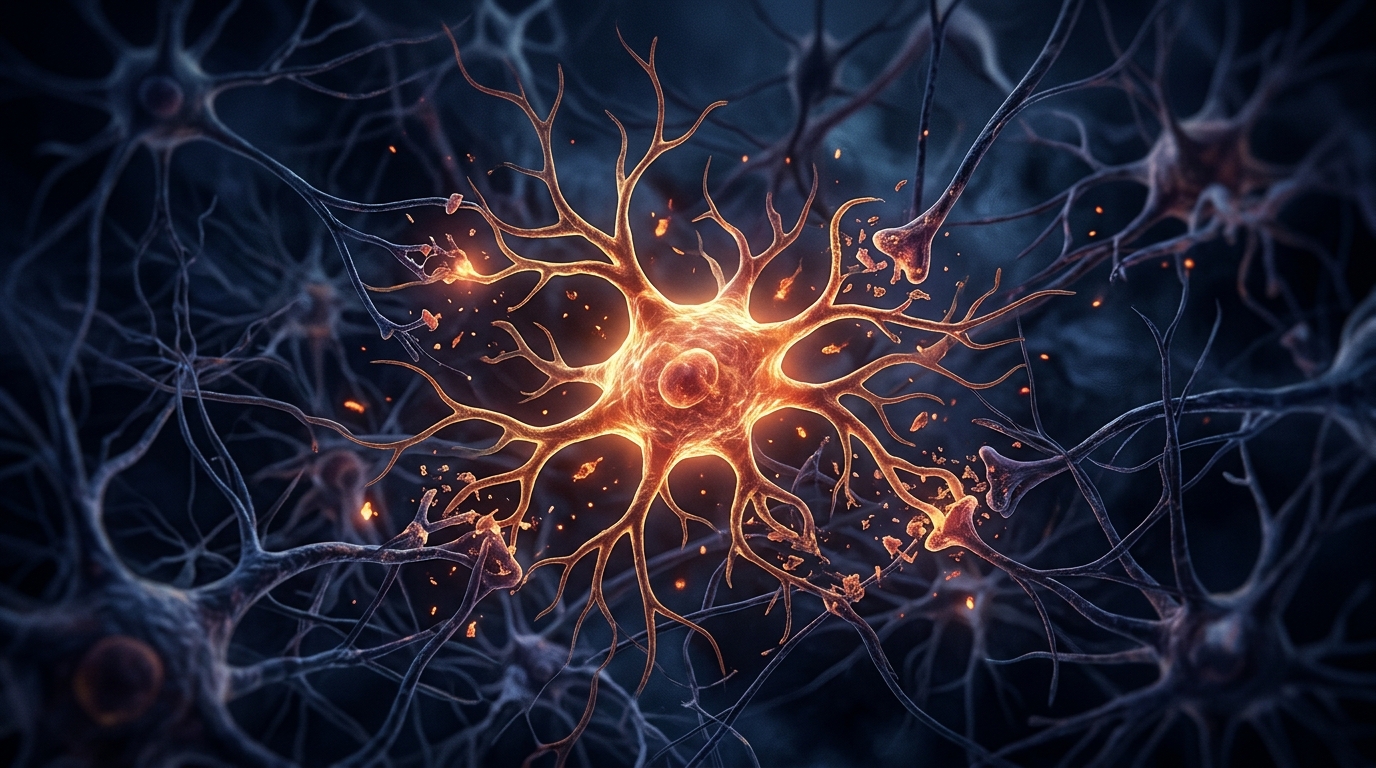
A single-cell study of mice with diffuse traumatic brain injury has identified a population of trauma-associated microglia that persists for weeks after the insult, reacts more violently to a later immune challenge, and contributes to lasting tissue damage.
Traumatic brain injury affects millions of people annually, and more than half of those individuals live with chronic symptoms, including cognitive dysfunction and neuropsychiatric complications. Inflammation after injury is thought to prime microglia, the resident immune cells of the brain, in ways that may alter normal immune responses and raise the risk of neurodegenerative pathology. The research team set out to map microglial profiles in the chronic phase after injury, both under an immune challenge with the bacterial molecule lipopolysaccharide, known as LPS, and after forced turnover of the microglial population using a CSF1R antagonist called PLX5622.
In the mouse model of diffuse injury, the researchers found that cortical inflammation persisted into the chronic phase and was associated with primed microglia and increased neuronal injury. That damage was measured through several markers: plasma neurofilament light chain, a blood-based indicator of neuronal damage, along with neuronal PTEN, NeuN and NSE in brain tissue. Thirty days after injury, a transient immune activation with LPS worsened the picture, exacerbating the morphological restructuring of cortical astrocytes and microglia and augmenting tissue injury.
Single-cell RNA sequencing of cortical cells at thirty days after injury revealed that roughly twenty percent of microglia had become trauma-associated microglia, abbreviated TRAM. These cells showed higher expression of differentially expressed genes that overlapped with two well-characterized microglial states: primed microglia, marked by genes such as Il1b, Cd74 and Cd14, and disease-associated microglia, marked by Apoe, Lyz2, Clec7a and Axl.
When the injured animals were challenged with LPS at thirty days after injury, the microglial response was amplified. The inflammatory program expanded while a heat shock regulatory response was reduced, indicating that the injured brain mounts a distorted version of a normal immune reaction. Notably, the single-cell RNA profile of microglia after LPS challenge in brain-injured mice was conserved within microglia from aged, twenty-four-month-old mice that received the same challenge.
With the immune challenge, trauma-associated microglia were highly reactive and displayed augmented pro-inflammatory profiles. These reactive TRAMs could be reduced by about fifty percent through forced turnover of microglia, which the researchers induced between seven and fourteen days after injury using the CSF1R antagonist. However, the intervention did not fully normalize the response. In the context of the overall reaction to LPS, forced turnover produced an intermediate activation profile rather than a complete return to baseline.
The partial restoration may be explained by a resistant subpopulation. A group of inflammatory microglia marked by low Csf1r expression, high Lgals3 expression and the MAC2 protein survived the PLX5622-mediated depletion, maintaining an inflammatory presence even after most microglia were eliminated. Nonetheless, other cell types benefited from the forced turnover.
The findings carry several caveats. The work was performed in mice with diffuse injury, and the chronic phase was examined at a single time point thirty days after insult. The LPS challenge is an experimental model of peripheral immune activation, not a direct replica of the infections or inflammatory events that people with brain injuries encounter. The forced turnover approach depletes and replaces microglia pharmacologically, a strategy that is not currently a clinical treatment. Whether the same trauma-associated microglial population exists in the human injured brain, and whether manipulating it would be safe or beneficial, remains to be established.
Subject of Research: Trauma-associated microglia in the chronic phase after traumatic brain injury
Article Title: Novel trauma-associated microglia influence immune reactivity and pathology in the chronic phase after traumatic brain injury
Article References: Wangler, L. M., Davis, A. C., Goodman, E. J., Bray, C. E., Lapurga, J. R., Gallagher, N. R., Luengo, R. A. M., Packer, J. M., Cummings, A., Kaiser, K. E., Khadkikar, P. P., Liu, T., El-Hodiri, H. M., Fischer, A. J., Harrington, C. A., Segal, B. M., & Godbout, J. P. (2026). Novel trauma-associated microglia influence immune reactivity and pathology in the chronic phase after traumatic brain injury. Acta Neuropathologica Communications. https://doi.org/10.1186/s40478-026-02468-8
Image Credits: AI Generated
DOI: 10.1186/s40478-026-02468-8
Keywords: traumatic brain injury, microglia, neuroinflammation, single-cell RNA sequencing, LPS challenge, CSF1R antagonist, PLX5622, astrocytes, neuronal injury, chronic inflammation, Novel, trauma-associated
News Source: Cassandra Pierce. (November 4, 2026). Trauma-associated microglia drive chronic inflammation after brain injury. Scienmag.