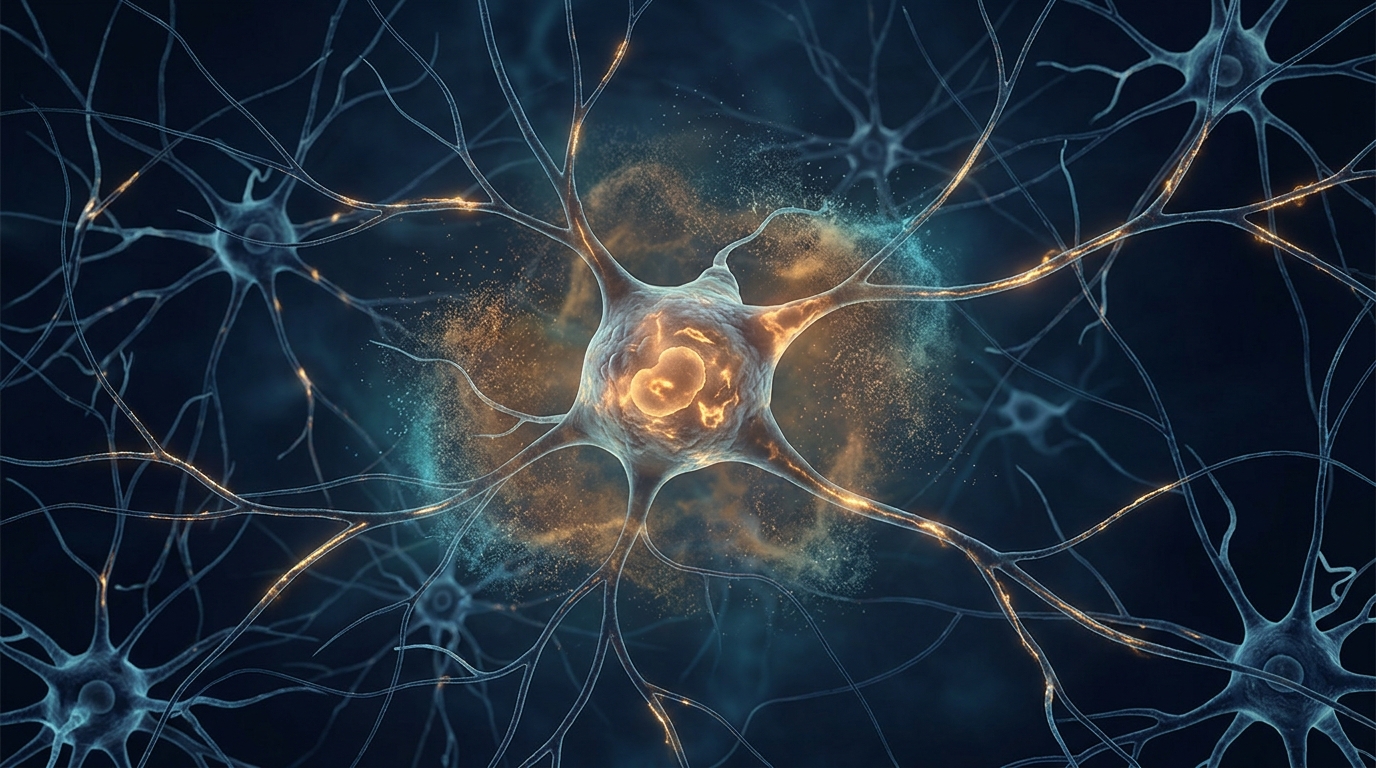
Why does pain fade in one patient and stubbornly persist in another after a seemingly identical injury? A new mini-review published in Anesthesiology and Perioperative Science argues that the answer may lie in a surprising cellular transformation: sensory neurons in the dorsal root ganglion, the clusters of nerve-cell bodies that relay pain signals from the body to the spinal cord, may enter a durable senescence-like state that locks in nociceptive output long after the original tissue damage has stabilized. The review, led by Jingxiu Huang, Mingkang Liu, Yingli Zhu, and Wangyuan Zou, synthesizes a decade of research and proposes that these altered neurons should be treated as a testable contributor to failed pain resolution rather than an established cause of chronic pain.
The authors searched PubMed, Web of Science, and Scopus for English-language articles published between January 2016 and July 2026, prioritizing original studies that identified the relevant cell type, assessed multiple senescence-associated features, incorporated temporal or functional measurements, or tested reversibility. Their central question was deliberately narrow: do durable senescence-like states in identified dorsal root ganglion sensory neurons contribute to failed pain resolution? The distinction matters because chronic pain remains common and difficult to treat, and after surgery the variation in the transition from acute to chronic pain is not explained by tissue injury alone. Recovery, the authors contend, should be viewed as an active biological process rather than the passive disappearance of an initiating stimulus.
The strongest direct evidence comes from a study by Donovan and colleagues, which combined histochemical, molecular, electrophysiological, behavioral, interventional, and human observations. In aged mice, the researchers found more dorsal root ganglion neurons with senescence-associated beta-galactosidase activity and increased expression of p21, p16INK4a, or interleukin-6. Peripheral nerve injury produced a time-dependent increase in these features in both young and aged animals, and the heterogeneous distribution of the markers suggested that only a subset of neurons was susceptible. Crucially, the marker-positive populations included high-firing and nociceptor-like cells, and senolytic treatment, drugs designed to clear senescent cells, reduced nociceptive behavior after nerve injury. Related neuronal markers also increased with age in human post-mortem dorsal root ganglia. By linking neuronal identity to several molecular features, altered firing, behavior, and an intervention, the study provides proof of principle for a functionally relevant senescence-like neuronal state.
Yet important limitations temper that conclusion. The markers did not converge uniformly within individual neurons, and the senolytic intervention was not restricted to dorsal root ganglion sensory neurons, meaning the behavioral improvement could have reflected effects on immune, glial, vascular, or stromal cells. The human samples were cross-sectional and could not link the neuronal features to pain history, temporal persistence, or recovery. The review is equally cautious about terminology: because mature sensory neurons are normally post-mitotic, expression of cell-cycle inhibitors such as p21 or p16INK4a cannot be interpreted as growth arrest the way it would be in a dividing fibroblast. Senescence-associated beta-galactosidase activity reflects increased lysosomal content but can occur outside classical senescence, and interleukin-6, though part of the senescence-associated secretory phenotype, is also released during conventional inflammation.
Independent support comes from single-cell analysis. The iPain atlas integrated dorsal root and trigeminal ganglion datasets from several injury models and identified senescence-associated transcriptional programs in a small subset of nociceptors. Senolytic treatment again reduced pain behavior in mice, and compatible signatures appeared in datasets from human chronic pain and painful diabetic neuropathy. These findings place the transcriptional signal within defined nociceptor populations and reinforce the idea of cellular heterogeneity. However, the authors caution that a computational score is not equivalent to multidomain phenotyping, and integrating experiments across studies may obscure differences in injury type, tissue processing, sampling time, and cellular recovery state.
To connect a durable neuronal state to failed pain resolution, the review outlines three candidate functional links. The first is persistent neuronal excitability. Donovan and colleagues identified high-firing and nociceptor-like cells within marker-positive populations, and human neuropathic dorsal root ganglion neurons also show spontaneous activity, indicating that persistent firing is not limited to rodent injury models. After injury, subtype-specific transcriptional programs remodel ion channels, receptors, and intracellular signaling. Epigenetic repression of the potassium-channel gene Kcna2 reduces potassium current and increases primary-afferent excitability, and a long non-coding RNA can maintain the same channel deficit. In isolated rat nociceptors, prolonged cAMP-PKA-CREB signaling produced transcription- and translation-dependent hyperexcitability twelve to twenty-four hours later, and after spared nerve injury, remodeling of N- and P/Q-type calcium channels increased calcium transients in central terminals of Trpv1-lineage afferents. These mechanisms can sustain abnormal membrane properties after nerve injury, but except for the Donovan study, none tested whether the affected neurons had a senescence-like phenotype.
The second candidate link is local neuron-glia communication. Sensory-neuron somata are surrounded by satellite glial cells and share the ganglion with resident macrophages, an arrangement that permits local signals to modify excitability without a new peripheral lesion. P2X7 receptor signaling in satellite glial cells contributes to persistent hypersensitivity in a surgical incision model, and single-cell analysis has identified metalloproteinase signaling in satellite glia as another pain-modifying pathway. In vivo calcium imaging adds an important qualification: inflammation and nerve injury increased the number of active satellite glial cells, but most glial calcium events were not time-locked to neuronal firing. Injured sensory neurons also release colony-stimulating factor 1, which induces a spinal microglial response, while within the ganglion, CXCL10/CXCR3 signaling increases neuronal excitability and neuropathic behavior. In an oxaliplatin study, disulfide HMGB1 traced from tissue-resident macrophages to Toll-like receptor 4 on a defined sensory-neuron subtype, and disrupting the pathway reduced mechanical allodynia. None of these studies tested whether a senescence-like neuron was the upstream source of glial activation.
The third link is metabolic dysfunction, which the authors describe as the closest experimental connection to pain resolution. Transient inflammation leaves mitochondrial and metabolic disturbances in sensory neurons after the initial hyperalgesia has resolved. Changes in ATP synthase c-subunit lysine methylation, oxidative phosphorylation, redox balance, and nicotinamide metabolism increased susceptibility to persistent hypersensitivity after a second inflammatory challenge, and correcting selected metabolic defects restored resolution. In a complementary study, suppressing mitochondrial pyruvate oxidation in the dorsal root ganglion caused persistent allodynia alongside widespread changes in gene expression, RNA splicing, and poly(A) tail length. These experiments show that metabolic reprogramming can outlast an initiating signal and alter neuronal output, though neither study classified the affected neurons as senescent. Satellite glial cells can even transfer mitochondria to neurons through myosin 10-dependent tunnelling nanotubes; blocking this transfer causes nerve degeneration and neuropathic pain, while mitochondria from healthy human satellite glia are protective.
What makes the review distinctive is its proposed testing ground: perioperative medicine. Because the preoperative baseline, timing of injury, perioperative exposures, and subsequent recovery can all be followed prospectively, surgery offers a uniquely observable model of failed recovery. After lung cancer surgery, serial measurements over twelve months identified complete, protracted, and incomplete recovery trajectories, with neuropathic symptoms most prominent in the incomplete-recovery group. In a larger abdominal-surgery cohort, persistently high acute-pain trajectories were associated with approximately twice the odds of chronic postsurgical pain, with subacute pain and early quality of recovery partly accounting for the association. The authors sketch an illustrative cohort assessing patients preoperatively, during postoperative days one through seven, and at approximately one, three, six, and twelve months, with repeated blood sampling to explore interleukin-6, CCL2, p16INK4a-associated transcripts, and cell-free mitochondrial DNA. They stress that such measurements may reflect systemic inflammation or stress in peripheral cells and cannot identify a senescence-like state in dorsal root ganglion neurons.
The bottom line is deliberately bounded. Aging and nerve injury can produce heterogeneous, multidomain senescence-like features in a subset of dorsal root ganglion sensory neurons, and the strongest evidence links these features to altered firing and to behavioral improvement after a non-neuron-specific senolytic intervention. But current evidence does not show that senescence-like dorsal root ganglion neurons are a common or necessary cause of persistent pain in humans, and no validated blood, cerebrospinal-fluid, or imaging measure of the state yet exists. Until experiments integrate neuronal identity, convergent state features, time, sensory function, recovery trajectory, and selective reversal within the same framework, the authors conclude that senescence-like dorsal root ganglion neurons should remain a focused, testable explanation for failed pain resolution, not a settled mechanism or an immediate therapeutic target.
Subject of Research: Senescence-like states in dorsal root ganglion sensory neurons as a proposed contributor to failed pain resolution
Article Title: Senescence-like states in dorsal root ganglion neurons and the failure of pain to resolve
Article References: Huang, J., Liu, M., Zhu, Y., & Zou, W. (2026). Senescence-like states in dorsal root ganglion neurons and the failure of pain to resolve. Anesthesiology and Perioperative Science, 4(1), Article 54. https://doi.org/10.1007/s44254-026-00202-0
Image Credits: AI Generated
DOI: 10.1007/s44254-026-00202-0
Keywords: chronic pain, dorsal root ganglion, cellular senescence, nociceptors, senolytics, neuron-glia signaling, mitochondrial dysfunction, neuronal excitability, perioperative medicine, pain resolution, satellite glial cells, single-cell transcriptomics
News Source: Beatrice Stafford. (November 4, 2026). Senescence-Like States in Sensory Neurons May Explain Why Pain Fails to Resolve. Scienmag.