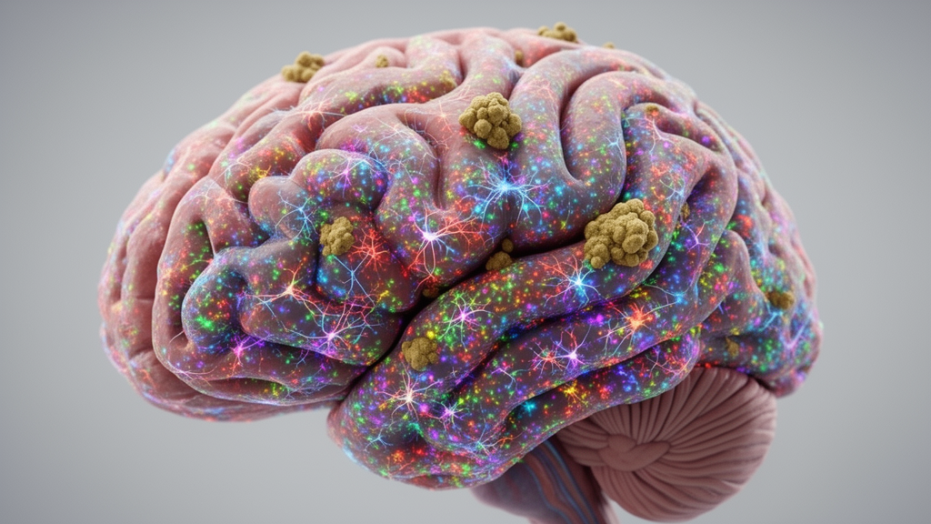
Spatial transcriptomic technologies are transforming Alzheimer’s disease research by mapping gene expression directly within intact brain architecture, bridging a century-old gap between anatomical pathology and molecular mechanisms. In a comprehensive review published in Molecular Neurodegeneration Advances, researchers led by Christina Huan Shi, Juan C. Piña-Crespo, Kevin Y. Yip, and Timothy Y. Huang synthesize how spatially resolved molecular methods are clarifying cellular reactions around classic disease hallmarks. Alzheimer’s disease has long been defined by the extracellular accumulation of beta-amyloid plaques and the intracellular aggregation of neurofibrillary tangles containing hyperphosphorylated tau. While these protein aggregates have historically served to stage disease onset in postmortem human brains, standard genomic profiling methods have struggled to preserve the physical orientation of cells relative to these lesions. Spatial transcriptomics allows investigators to measure transcriptomic changes directly in native tissue microenvironments across both human patient samples and murine experimental models, offering fresh insight into regional vulnerability, resilience, and microenvironmental disruption.
The study of Alzheimer’s disease neuropathology originated more than a century ago when Alois Alzheimer detailed memory loss and atypical brain lesions in his patient Auguste Deter at a 1906 psychiatric conference in Tübingen. Although his presentation initially drew little discussion and was initially deemed unsuitable for meeting proceedings, a concise report published in 1907 established the foundation for modern dementia investigation. For decades, traditional bulk RNA sequencing successfully cataloged broad transcriptomic alterations across brain regions, yet it pooled together disparate cell types and lost fine spatial resolution. Single-cell and single-nucleus RNA sequencing subsequently revolutionized the identification of distinct cellular states, but these methods require tissue dissociation, completely severing cells from their native anatomical coordinates and surrounding pathological landmarks. Spatial transcriptomics overcomes these limitations by capturing messenger RNA expression in situ, enabling researchers to correlate transcriptional dynamics directly with local amyloid and tau burden in intact sections.
Current-era spatial transcriptomics encompasses distinct platform classes that each balance resolution against transcriptomic breadth. Array-based next-generation sequencing approaches capture messenger RNA across patterned slides, spots, beads, or nanoballs. The widely adopted 10x Genomics Visium platform evaluates spots fifty-five micrometers in diameter containing approximately one to ten cells, while newer iterations such as Visium HD feature two-micrometer square barcoded capture areas. Related barcode methods like Slide-seq, Slide-seqV2, and deterministic barcoding in tissue for spatial omics sequencing deploy microfluidic channels or bead arrays spanning ten to fifty micrometers. Stereo-seq incorporates patterned arrays of DNA nanoballs roughly 220 nanometers in diameter with center-to-center distances between 500 and 715 nanometers. Because individual nanoballs yield sparse transcript capture, raw Stereo-seq readouts are routinely grouped into larger computational bins, such as twenty-five-micrometer bins comprising fifty-by-fifty nanoballs, to maintain sufficient transcript detection sensitivity across broad tissue expanses.
In contrast to array-based sequencing, imaging-based platforms profile transcripts in situ with subcellular precision through in situ hybridization or in situ sequencing. Commercial systems including CosMx from NanoString, Multiplexed Error-Robust Fluorescence In Situ Hybridization on the MERSCOPE platform from Vizgen, and Xenium from 10x Genomics utilize complementary fluorescent probes or optical decoding to detect spatial coordinates of target transcripts. CosMx achieves subcellular precision with pixel edges down to 120 nanometers for panels typically ranging from 1,000 to 6,000 genes, while MERSCOPE achieves 100-nanometer pixel edges across gene panels of up to 1,000 targets, and Xenium resolves targets at approximately 200 nanometers across panels reaching 5,000 genes. While imaging platforms pinpoint transcript locations with high spatial fidelity, they generally monitor preselected gene panels rather than unbiased whole transcriptomes. Consequently, investigators frequently integrate spatial transcriptomic datasets with matched single-cell RNA sequencing data, computationally imputing whole-transcriptome measurements across single-cell spatial maps or inferring the spatial distribution of single-cell clusters across tissue architecture.
Applying these platforms to Alzheimer’s disease brain tissue requires careful experimental accommodation for sample preservation and histological compatibility. Human postmortem tissue is predominantly archived as formalin-fixed paraffin-embedded blocks, where chemical crosslinking and temporal decay degrade RNA integrity. Modern platforms address this limitation through specialized probe hybridizations tailored to fragmented transcripts, such as probe-based chemistry in Visium and decrosslinking protocols in MERFISH. Furthermore, resolving pathological microenvironments requires multiplexed histopathological staining on the very tissue sections undergoing sequencing. Immunofluorescence staining using antibodies such as 6E10, MOAB-2, 4G8, or 6F/3D, alongside chemical dyes like Amylo-Glo and Congo red derivative X-34, allows researchers to locate amyloid plaques. Tau pathology is predominantly mapped using the AT8 antibody, which targets serine 202, threonine 205, and serine 208 phosphoepitopes characteristic of paired helical filaments. Immunofluorescence can be executed prior to transcript capture in array platforms like Visium and Stereo-seq or following transcriptomic runs in imaging systems like CosMx and Xenium.
Computational processing of spatial transcriptomic data introduces unique challenges, beginning with cell segmentation in densely packed brain tissue. Defining accurate boundaries between cells or nuclei is often confounded by dense neuropil, overlapping cell bodies, and significant optical background from pathology. Notably, nuclear counterstains like DAPI can non-specifically label amyloid plaques, creating false-positive signals in regions with advanced pathological burden. To improve segmentation, analysts deploy combinatorial staining strategies, such as combining DAPI with polyT, alongside machine learning architectures refined through human-in-the-loop manual boundary annotations. Following segmentation, transcript counts are normalized using total counts, variance-stabilizing transformations, or segmented cell volumes, which prove particularly advantageous when evaluating constrained gene panels. Tissue sections are routinely registered to anatomical coordinate systems, such as the Allen Mouse Brain Common Coordinate Framework, or segmented de novo through spatial clustering algorithms like BayesSpace to distinguish neocortical layers and white matter tracts based on localized gene expression signatures.
Spatial transcriptomic investigations have illuminated molecular mechanisms underlying regional vulnerability and selective resilience across Alzheimer’s disease progression. In human hippocampal tissue, heightened amyloid plaque accumulation in the CA1 sector coincides with profound pyramidal neuron loss alongside localized microglia and astrocyte accumulation, whereas excitatory neurons in CA3 and CA4 regions exhibit relative resistance to degeneration. Stereo-seq combined with single-nucleus sequencing in human hippocampus revealed elevated synaptic pruning machinery in the fimbria and enhanced metabolic gene activity across the cornu ammonis. Conversely, spatial profiling in human primary visual cortex identified a resilient excitatory neuron subset in layer four co-expressing RORB, CUX2, and EYA4 that displays upregulated KCNIP4 expression. Because KCNIP4 encodes a potassium channel-interacting protein that dampens neuronal excitability, functional murine testing confirmed that its elevation suppresses activity-dependent immediate early genes such as Arc and c-Fos, suggesting that intrinsic excitability suppression may restrict trans-synaptic tau propagation.
High-resolution spatial profiling has also unraveled localized, multicellular responses within the immediate microenvironment surrounding amyloid plaques. Early spatial transcriptomic studies in humanized APPNL-G-F mouse models uncovered fifty-seven plaque-induced genes involved in complement activation, lysosomal function, and oxidative stress, which in situ sequencing subsequently localized primarily to reactive microglia and astrocytes. Around amyloid cores, disease-associated microglia assemble into an intimate inner protective envelope, encircled by an outer perimeter of disease-associated astrocytes and oligodendrocyte lineage cells. Genetic modifiers critically shape these localized glial envelopes. In 5xFAD mice crossed with human APOE4 knock-in alleles, spatial analysis revealed exacerbated upregulation of reactive microglial programs and synaptic pruning signatures compared to APOE3 counterparts.
Spatially resolved cell-cell interaction algorithms, including CellPhoneDB, NICHES, and StereoSiTE, demonstrate that pathological niches systematically rewire local intercellular communication networks. In human dorsolateral prefrontal cortex, regions with elevated amyloid deposition exhibit marked upregulation of ligand-receptor pairs governing immune activity, such as CLU-TREM2, PLXNA1-TREM2, APOE4-LDLR, and GFAP-APLNR, alongside extracellular matrix signaling. Conversely, transcriptomic signatures indicative of synaptic degeneration and receptor-ligand decoupling predominate in tissue domains displaying minimal amyloid burden, suggesting that degenerative processes may initiate prior to widespread plaque deposition. In human hippocampus, Stereo-seq datasets highlighted enriched inflammatory crosstalk between reactive microglia and disease-associated astrocytes within the fimbria, notably via SPP1-ITGB1 and MIF-CD44 signaling pathways, while NECTIN-mediated synaptic adhesion signaling is progressively downregulated in Down syndrome and advanced Alzheimer’s cases.
Temporal analyses derived from spatial transcriptomics further indicate that disease progression does not follow monotonic gene expression shifts, but instead exhibits distinct stage-dependent regulatory phases. For instance, the neuroprotective gene PSAP undergoes early upregulation in prefrontal cortex before undergoing pronounced downregulation in advanced disease stages. Similarly, transient upregulation of synaptic genes including SNAP25, GABRA1, GRIN2B, and SYT1 is detected in neurofibrillary tangle-bearing neurons, reflecting compensatory neuroprotective efforts before structural disconnection. Looking toward future applications, integrating spatial transcriptomics with spatial epigenomics, proteomics, and longitudinal imaging will be critical for reconstructing multi-omic disease trajectories. As spatial profiling advances toward true subcellular resolution with whole-transcriptomic coverage, anchoring localized cellular programs to fluid biomarkers and neuroimaging signatures will accelerate patient stratification and guide the development of stage-specific therapeutic interventions.
Subject of Research: Spatial transcriptomics applications and discoveries in Alzheimer's disease neuropathology
Article Title: Spatial transcriptomics in Alzheimer's disease: technologies, challenges and discoveries
Article References: Shi, C. H., Piña-Crespo, J. C., Yip, K. Y., & Huang, T. Y. (2026). Spatial transcriptomics in Alzheimer's disease: technologies, challenges and discoveries. Molecular Neurodegeneration Advances, 2(1), Article 30. https://doi.org/10.1186/s44477-026-00036-x
Image Credits: AI Generated
DOI: 10.1186/s44477-026-00036-x
Keywords: Alzheimer's disease, spatial transcriptomics, amyloid beta, neurofibrillary tangles, single-cell RNA-seq, microglia, astrocytes, Visium, Stereo-seq, CosMx, MERFISH, Xenium, neurodegeneration, cell segmentation, cell-cell interactions, hippocampus, disease-associated microglia
News Source: Cassandra Pierce. (November 21, 2026). Spatial Transcriptomics Resolves Alzheimer Pathology and Cellular Changes Across Intact Brain Tissue. Scienmag.