Sound has become an unlikely tool for sculpting living tissue in the laboratory. In a study published in BMC Methods, researchers describe a workflow that uses low-frequency acoustic waves to gather thousands of human bone marrow–derived mesenchymal stromal cell spheroids into dense circular rings inside a three-dimensional fibrin hydrogel, and then to stimulate those same constructs with daily acoustic pulses. The result was not merely a change in geometry. RNA sequencing showed that both the assembly step and the subsequent stimulation left measurable fingerprints on the cells’ gene expression, pointing to shifts in stress regulation, cell–cell communication, matrix remodeling and mechanotransduction.
The method, built around a platform the authors call Sound Induced Morphogenesis, or SIM, addresses a gap that has persisted as acoustic biopatterning has grown in popularity. Acoustic fields can move cells and multicellular aggregates without touching them, which makes them attractive for three-dimensional culture and mechanobiology. Yet, according to the team, standardized and reproducible protocols that combine sound-guided assembly with controlled acoustic stimulation and downstream molecular analysis have been lacking. Their paper, a methods article rather than a clinical study, lays out such a protocol in detail, from hydrogel rheology to sequencing bioinformatics, and reports the transcriptomic consequences observed in a proof-of-concept experiment.
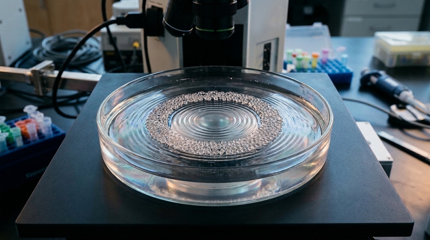
The biological raw material was hBMSC spheroids, each containing roughly 260 cells, produced in standard microwell plates from cells originally obtained from a single 58-year-old male donor undergoing spinal surgery, with written informed consent and ethical approval. About 2.8 × 10⁴ spheroids were collected, mixed into a fibrinogen-based precursor solution together with thrombin, and poured into shallow circular frames roughly 15 millimeters across. When the SIM device vibrated the liquid surface at 36 hertz for just two to five seconds, the spheroids rapidly migrated into a well-defined annular pattern. Gelation was then triggered by incubation at 37 °C, locking the arrangement in place as the fibrin clot formed.
Getting that to work reliably required the hydrogel precursor to behave properly under vibration. Rheological testing with a parallel-plate rheometer showed a complex viscosity of around 400 millipascal-seconds that stayed linear over five minutes, and an oscillatory time sweep indicating a very fast sol-to-gel transition that formed a stable gel. These measurements defined the operational window during which acoustic patterning could be applied before the matrix stiffened. The kinetics of sound-driven assembly in fibrin and gelatin-based precursors is fast, the authors note, taking less than five seconds, so timing is everything.
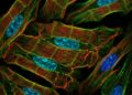
Three experimental groups were compared over seven days of culture in chondropermissive medium. Dispersed spheroids, labeled D, were embedded randomly in the fibrin. Assembled spheroids, labeled A, were patterned into rings. A third group, A + S, was assembled and then exposed to additional acoustic stimulation at 100 hertz for one hour per day, applied through the closed Petri dish lid to maintain sterility, with four replicates in each group. Imaging told a clear story. Dispersed constructs showed a disordered, diffuse distribution with weak and random F-actin staining, while assembled constructs formed compact rings with strong F-actin signal and increased nuclear density, consistent with enhanced cell condensation and cytoskeletal alignment. Crucially, the stimulated group kept its circular configuration throughout the week, showing that the daily acoustic pulses preserved structural integrity without disrupting the pattern.
Quantitative image analysis backed up the visual impression. Using a radial profile plugin in ImageJ, the team measured normalized integrated intensity as a function of distance from the construct center. Dispersed samples produced a low, broadly uniform profile typical of scattered particles, whereas assembled samples showed a distinct, narrow intensity peak at the ring’s radial position. That peak persisted in the stimulated group, confirming the stability of the organization over time. The authors suggest the ring geometry is biologically meaningful because it concentrates spheroids into a high-density annulus while maintaining a lower-density surrounding zone, distributing mechanical stress isotropically and promoting balanced radial diffusion of nutrients and paracrine factors.
The transcriptomic readout came from RNA sequencing of the three conditions, with differential expression analyzed using DESeq2 and validation by RT-qPCR on six selected genes. Unsupervised hierarchical clustering separated the samples into three groups of genes with distinct expression profiles. Comparing assembled with dispersed spheroids, 11 genes were upregulated and 6 were downregulated. Adding acoustic stimulation upregulated a further set of 8 genes relative to assembled but unstimulated spheroids. Among the downregulated genes in the assembled group were CXCL6, which encodes a chemokine involved in inflammation, cell migration and proliferation, and SOD2, which encodes the mitochondrial antioxidant enzyme superoxide dismutase 2. The authors hypothesize that both changes reflect the local density enhancement: when cell–cell communication improves in the absence of an inflammatory stimulus, chemokine expression may be less needed, and oxidative stress and stress fiber formation are known to decrease when cells reach confluence, reducing the demand for superoxide scavenging.
The upregulated side of the assembly signature included ITGA8, encoding the alpha-8 subunit of an integrin that binds fibronectin, which the authors interpret as evidence that cells sense the initial assembly process through integrin-mediated adhesions. Also elevated were TMEM64, implicated in ATP-dependent metabolic activity, and CHRDL1, a gene reported to influence topographic projection and retinal angiogenesis, which the authors read as a hint that acoustic stimulation can act as a guidance signal for cells. Both TMEM64 and CHRDL1 are described as negative regulators of differentiation and positive promoters of proliferation, aggregation and extracellular matrix formation. The stimulated group, meanwhile, switched on a distinct set of transcripts, many of them long noncoding RNAs or sequences of unknown function, alongside protein-coding genes tied to osteogenic differentiation and mechanoresponses, including IBSP, SNX22, MMP13 and DES, the gene for the intermediate filament protein desmin, as well as MXRA5, an adhesion proteoglycan involved in matrix remodeling.
The mechanistic picture the authors sketch is that repeated acoustic forces travel through the fibrin matrix to integrin receptors established during the initial assembly, are transduced intracellularly through the cytoskeleton, and converge on transcriptional programs associated with matrix formation and early osteogenic activation. RT-qPCR confirmed the significant downregulation of CXCL6 and SOD2 in assembled versus dispersed spheroids, with both genes trending non-significantly back upward when assembled spheroids were further stimulated; similar non-significant trends appeared for CXCL8 and IBSP. The team acknowledges that the 100-hertz, one-hour-per-day regimen was chosen based on prior work showing that low-frequency vibrations between 80 and 120 hertz transmit mechanical cues through soft hydrogels while preserving construct integrity, and that alternative frequencies, amplitudes or temporal patterns could produce different cellular outcomes. Future studies, they write, will systematically compare stimulation regimes to map the frequency- and dose-dependence of acoustic mechanoactivation in these cells.
What distinguishes this work from earlier acoustic patterning demonstrations, the authors argue, is the combination of physical patterning with molecular readout. Previous studies from the same group used sound-guided assembly to organize endothelial cells and pericytes into capillary-like structures, accompanied by broad protein remodeling, and to build an in vitro model of annulus fibrosus innervation in which spatial proximity between tissue units drove functional crosstalk and axon guidance. Here, the focus shifts to stem cells and their transcriptomes. The team also contrasts sound-guided assembly with alternative patterning strategies such as photopatterning, scaffolding, bioprinting or magnetic assembly, which they note rely on surface modification, physical confinement or chemical gradients that introduce additional variables. Acoustic assembly, by contrast, works through transient pressure gradients and hydrodynamic flows that condense spheroids contactlessly without changing the biochemical composition of the matrix. The RNA sequencing data have been deposited in the NCBI Gene Expression Omnibus under accession GSE319241, and the authors position the workflow as a transferable framework for mechanobiology research and engineered tissue model development, adaptable to other cell types and hydrogels, and potentially able to complement or reduce reliance on growth factors in tissue engineering strategies.
Subject of Research: Sound-guided acoustic assembly and stimulation of human mesenchymal stromal cell spheroids in 3D fibrin hydrogels and its effects on gene expression
Article Title: Control of human mesenchymal stromal cell gene expression levels by sound-guided assembly and stimulation
Article References: Della Bella, E., Daou, F., Cianciosi, A., Ma, J., Tognato, R., Alini, M., Cochis, A., Rimondini, L., Stoddart, M. J., & Serra, T. (2026). Control of human mesenchymal stromal cell gene expression levels by sound-guided assembly and stimulation. BMC Methods, 3(1), Article 15. https://doi.org/10.1186/s44330-026-00070-x
Image Credits: AI Generated
DOI: 10.1186/s44330-026-00070-x
Keywords: acoustic patterning, mesenchymal stromal cells, spheroids, fibrin hydrogel, mechanotransduction, RNA sequencing, gene expression, 3D cell culture, mechanobiology, Sound Induced Morphogenesis, tissue engineering, RT-qPCR
News Source: Juliet Wilcox. (November 5, 2026). Sound waves arrange stem cell spheroids into rings and reshape their gene expression. Scienmag.