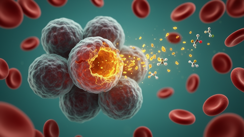
Ferroptosis, an iron-dependent form of cell death driven by runaway lipid peroxidation in cellular membranes, has long been difficult to detect reliably, particularly inside living organisms. Now a large multi-institution team led by Jianping He and Zachary T. Schafer, publishing in Nature Cell Biology, reports the discovery of an unexpected molecular signature that appears when cells undergo this type of death: a modified form of the transferrin receptor 1 that resists the reducing chemicals normally used to break apart protein dimers during laboratory analysis. The researchers call this structure the reducing reagent-resistant dimer, or 3R dimer, and they show that it can be detected not only in cultured cells but also in tissue lysates and in extracellular vesicles isolated from the blood of mice and human patients.
The discovery began with a routine western blot observation. TfR1 is a type II transmembrane protein that normally forms homodimers through disulfide linkages; under non-reducing conditions most of the protein appears dimeric, while standard reducing conditions collapse it to the monomeric form. When the authors treated 786-O kidney cancer cells or MDA-MB-231 breast cancer cells with RSL3, an inhibitor of the ferroptosis-suppressing enzyme GPX4, a high molecular weight band of TfR1 appeared even under reducing conditions. The band was confirmed with two independent antibodies, and short hairpin RNA targeting TFRC, the gene encoding TfR1, lowered both the mystery band and monomeric TfR1, ruling out a non-specific artifact. In cells engineered to express HA-tagged TfR1, the band was also detected with an anti-HA antibody, and mass spectrometry of immunoprecipitated material confirmed that TfR1 protein was present and enriched after RSL3 treatment.
What made the band remarkable was its stubbornness. Extending heat exposure or increasing the concentration of the thiols routinely used in western blotting, including beta-mercaptoethanol, dithiothreitol and tris(2-carboxyethyl)phosphine, only marginally disrupted the structure. Mutational analysis pinpointed the chemistry: TfR1 carries four cysteine residues near its transmembrane domain, and mutating cysteines 62 and 67 diminished dimer formation in response to RSL3, while mutating cysteines 89 and 98, or all four at once, hindered dimer formation under both reducing and non-reducing conditions. The researchers also found that the sodium dodecyl sulfate concentration in the sample buffer affected detection, probably because TfR1 is heavily glycosylated, and they settled on 3 percent SDS for subsequent experiments. With that, the team had named and characterized a new biochemical entity: the 3R dimer.
The next question was whether the 3R dimer was specific to ferroptosis or a generic stress response. The answer, across an extensive panel of experiments, was specificity. All four known classes of ferroptosis-inducing compounds triggered dimer accumulation: erastin, which targets the system Xc− cystine importer; RSL3 and ML210, which covalently inhibit GPX4; FIN56, which disrupts the mevanonate pathway; and FINO2, which promotes iron-dependent oxidation. Cystine starvation and genetic knockout of GPX4 in HT-1080 and 293T cells, followed by withdrawal of the ferroptosis inhibitor ferrostatin-1, produced the same result. Ferroptosis-sensitive cell lines including RCC4, RCC10 and 4T1 accumulated the dimer after RSL3 exposure, whereas ferroptosis-resistant MDA-MB-436 and A549 cells did not unless they were sensitized by inhibition of the ferroptosis suppressor protein FSP1. Ferroptosis inhibitors, including ferrostatin-1, liproxstatin-1, the iron chelator deferoxamine and the antioxidant Trolox, all prevented the dimer from rising.
Equally telling was what did not raise the signal. Etoposide-induced apoptosis in Jurkat cells, staurosporine treatment, TNF-driven necroptosis in L929 cells, LPS and nigericin-induced pyroptosis in THP-1 monocytes, glucose-starvation-induced disulfidptosis in SLC7A11-high cells, and elesclomol-induced cuproptosis all killed cells robustly without elevating the 3R dimer. Even hydrogen peroxide, which causes extensive oxidative cell death, failed to induce the dimer unless iron chemistry was engaged; tert-butyl hydroperoxide, an organic peroxide that can kill by ferroptosis, did induce it. Inhibiting glutathione synthesis with buthionine sulfoximine alone was insufficient, but adding ferric ammonia citrate alongside it drove both ferroptosis and dimer formation. Proteotoxic stress from MG132 and endoplasmic reticulum stress from tunicamycin also left the dimer untouched. The 3R dimer, in short, tracks lipid peroxidation tied to the ferroptotic program rather than cell death in general.
Mechanistically, the team turned to a cell-free system. Isolated cellular membranes treated with ferrous sulfate, which drives the Fenton reaction, showed a marked increase in lipid peroxidation and a corresponding rise in the 3R dimer, an effect reversed by deferoxamine, radical-trapping agents or the detergent Triton X-100. The four-cysteine mutant abrogated dimer elevation even under robust peroxidation, confirming the structural requirement. The authors then suspected the bifunctional aldehydes malondialdehyde and 4-hydroxynonenal, well-known byproducts of lipid peroxidation that covalently modify proteins. Direct application of either aldehyde to isolated membranes promoted 3R dimer formation without any ferroptosis inducer, and the R651A mutation, which blocks transferrin binding, did not affect dimer elevation, showing the process is independent of the transferrin interaction. Related receptor family members, TfR2 and NAALADL2, also formed reducing reagent-resistant dimers upon ferroptosis induction.
In a striking proof of principle, the team showed that the dimer-forming capacity could be transferred to an unrelated protein. ST6GAL1, a Golgi and endoplasmic reticulum glycosyltransferase of the same type II membrane architecture, does not form a 3R dimer on its own. But chimeric proteins combining domains of TfR1 and ST6GAL1 formed the resistant dimer in response to RSL3 or ferrous sulfate treatment only when they carried the TfR1 extracellular domain. This suggests the aldehyde-mediated crosslinking requires preexisting proximity of two monomers, meaning reducing reagent-resistant dimers are not created from scratch during ferroptosis but stabilized from dimers that already exist at baseline. The finding provides a design template for building ferroptosis sensors into other membrane proteins.
The in vivo experiments carried the marker into whole organisms. In a mouse model of multiorgan dysfunction syndrome induced by intraperitoneal iron sulfate injection, the 3R dimer rose in liver and kidney tissue lysates, and its abundance correlated positively and linearly with malondialdehyde levels across animals. Pretreating mice with ferrostatin-1, UAMC-3203 or liproxstatin-1 blocked the organ damage, the lipid peroxidation and the dimer induction. A tamoxifen-inducible, hepatocyte-specific Gpx4 knockout model produced the same pattern: elevated tissue damage, elevated malondialdehyde and elevated 3R dimer in the liver. As a specificity control, the authors used a urinary tract infection model driven by uropathogenic Escherichia coli, which kills bladder cells through pyroptosis; despite detectable cleaved GSDMD and even increased TfR1 expression, no rise in the 3R dimer appeared.
Perhaps the most clinically consequential finding involves extracellular vesicles. Because TfR1 is trafficked to these membrane-bound particles, and because cells undergoing lytic death release them in elevated quantities, the team asked whether ferroptotic cells shed vesicles carrying the 3R dimer. In cultured Jurkat cells, only ferroptosis induction, not apoptosis, necroptosis or pyroptosis, produced vesicles enriched in the dimer, with normalization to the vesicle markers ALIX and TSG101 confirming the result was not an artifact of vesicle number. In the mouse iron-overload model, plasma vesicles showed increased 3R dimer that was blocked by ferroptosis inhibitors and correlated with plasma malondialdehyde. The team then examined two human settings: five glioblastoma patients treated in a clinical trial with iron nanoparticles plus ascorbic acid all showed strong post-treatment induction of the 3R dimer in plasma vesicles alongside elevated malondialdehyde, and in ten inflammatory bowel disease patients selected for declining malondialdehyde levels upon flare resolution, the dimer present in vesicles during disease flare fell appreciably in every case after treatment.
The authors are candid about limitations. Detecting the 3R dimer requires SDS–PAGE and immunoblotting, its sensitivity in extracellular vesicles may be lower than in tissue samples, and patient-to-patient heterogeneity in baseline lipid peroxidation and TfR1 expression will complicate interpretation. The exact amino acid residues involved in the aldehyde-mediated crosslink remain unidentified. Even so, the marker offers practical advantages: it can be detected with multiple commercially available antibodies or with tagged versions of TfR1, it exploits the near-ubiquitous expression of TfR1, and it provides a signal visually distinct from the monomeric protein. By offering a durable, blood-accessible readout of ferroptotic cell death, the 3R dimer gives researchers a new tool for identifying cell death modalities, probing ferroptosis in disease, and screening for drugs that modulate this increasingly consequential form of regulated cell death.
Subject of Research: A reducing reagent-resistant TfR1 dimer as a specific biomarker for detecting ferroptosis in cells, tissues and extracellular vesicles
Article Title: The rise of reducing reagent-resistant TfR1 dimer marks ferroptosis
Article References: He, J., Kelleher, M., Veeckmans, G., Zhang, Y., Reynolds, L., Tanczos, H. M., Huang, Z., McLaughlin, B. P., Martinez Molina, M. A., Lightburn, A., Zamarro, M., Huang, W., Owusu, S. B., Walravens, M., Willemart, C., Hassannia, B., Pope, L. E., Gager, C., Vaughan, A. J., … Schafer, Z. T. (2026). The rise of reducing reagent-resistant TfR1 dimer marks ferroptosis. Nature Cell Biology. https://doi.org/10.1038/s41556-026-02107-3
Image Credits: AI Generated
DOI: 10.1038/s41556-026-02107-3
Keywords: ferroptosis, transferrin receptor 1, lipid peroxidation, biomarker, extracellular vesicles, cell death, GPX4, malondialdehyde, iron overload, western blotting, multiorgan dysfunction syndrome, inflammatory bowel disease
News Source: Drew Townsend. (October 28, 2026). Scientists discover a durable molecular fingerprint of ferroptosis in cells and blood. Scienmag.