Biomechanics researchers are increasingly combining imaging, mechanical testing, computational modeling, biomaterials and advanced manufacturing to understand how healthy and pathological tissues respond to mechanical stimuli, and how their interactions with medical devices can be optimized. A new editorial in Frontiers in Bioengineering and Biotechnology gathers the fruits of that effort, drawing on work presented at the Annual Meeting of the Italian Chapter of the European Society of Biomechanics, held in Pescara from 3 to 4 October 2024. The associated Research Topic collects six studies that, despite spanning very different organ systems, share a common quantitative ambition: to link mechanical behavior with biological or clinical function in ways that could eventually improve diagnosis and treatment.
The editorial, authored by Cristina Falcinelli of the Department of Engineering and Geology at the University G d’Annunzio Chieti-Pescara, emphasizes that integrating these approaches is important for capturing the complex relationships between tissue structure, mechanical properties and function. Progress in imaging, experimental testing, computational modeling, biomaterials and advanced manufacturing, the author notes, has opened new opportunities to characterize biological systems and translate that knowledge into improved diagnosis and treatment. The six contributions address dental and craniofacial systems, the respiratory system, the urinary system and the cardiovascular system, and together they illustrate how innovative approaches can give a more in-depth knowledge of healthy and pathological tissues and support the development of clinically relevant solutions.
Three of the studies focus on dental and craniofacial questions, and they operate at three different scales: the cell, the tissue and the organ. At the cellular level, Putame and colleagues developed a bioreactor platform for applying controlled intermittent stretching to human periodontal ligament stem cells. By characterizing the deformation of the cell substrate and comparing responses across stretching protocols, the team showed that greater daily cyclic exposure was associated with cell alignment and increased expression of osteogenic and periodontal-ligament-related genes. Notably, adipose-derived stem cells did not show the same response under the tested conditions, a contrast the editorial highlights as underscoring the importance of tissue-specific mechanobiology.
At the tissue level, Stróżyk turned to digital image correlation, or DIC, to map the surface displacement of the temporalis muscle in vivo during unilateral chewing. In a preliminary study of eight healthy men, the measurements revealed distinct regional kinematic patterns and showed that local motion varied with chewing side and food texture. The editorial argues that these findings support the use of non-contact, full-field measurements to investigate muscle function during physiological tasks.
At the organ level, Becker and colleagues compared subject-specific mandibular finite element predictions with experimental measurements, using eight different density-elasticity relationships. The study showed that predictions varied substantially depending on which density-elasticity relation was used, and that none of the relationships consistently reproduced the experimental response, particularly the local surface strain. The editorial frames this finding as a cautionary tale: constitutive assumptions affect model predictions, and experimental validation and sensitivity analysis remain essential. In other words, even sophisticated patient-specific models of the jawbone can produce materially different answers depending on how the relationship between bone density and elastic modulus is specified.
Biomechanical approaches were also applied beyond the craniofacial domain, to respiratory, urinary and cardiovascular systems. Huang and colleagues combined tensile tests of human lung bullae and parenchyma with computed tomography-derived, geometry-specific finite element models. Although the bullae showed higher tensile strength than adjacent parenchyma, the computed stress concentrations at the bulla apex offer a plausible mechanical explanation for the clinical tendency toward apical rupture. The result is a striking example of how geometry and local mechanics can matter more than bulk material strength: the computed stress concentrations at the apex, rather than the measured tensile strength alone, provide the mechanical account of why ruptures tend to occur there.
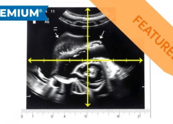
Liu and colleagues paired transvaginal ultrasound with peristaltic analysis to quantify ureteral wave characteristics in healthy women. Their pilot study demonstrated the feasibility of non-invasive measurement of dynamic parameters, including wave speed and transmission distance, and provides a basis for testing the method in patients with ureteral disorders. The editorial notes that this study extends the quantitative approach toward functional, non-invasive assessment. Because the technique was demonstrated in healthy women, its value in clinical populations remains to be established, but the pilot establishes the measurement foundation on which such patient studies can now be built.
Finally, Mazzoli and colleagues combined statistical shape modeling and computational fluid dynamics to examine how aortic morphology affects the watershed zone during veno-arterial extracorporeal membrane oxygenation, or VA-ECMO, support. Their simulations indicate that both aortic geometry and ECMO flow level influence the zone’s position and distribution, highlighting anatomical variability as a factor in hemodynamic assessment.
Across these systems, the studies investigate how tissue properties, geometry, loading and measurement choices influence the observed mechanical or biological response. The mandibular study highlights the sensitivity of model predictions to constitutive assumptions; the lung and VA-ECMO studies show that anatomy can influence local mechanics and flow; and the dental cell and muscle studies link mechanical stimulation or movement patterns to biological function. The methods span mechanical testing, bioreactor experiments, DIC, ultrasound-based motion analysis, CT-based geometric reconstruction, subject-specific finite element analysis, statistical shape modeling and computational fluid dynamics. The editorial characterizes the six contributions as sharing a quantitative approach to linking mechanical behavior with biological or clinical function.
Looking ahead, the editorial argues that future advancements will depend on validation in larger cohorts and clinically relevant conditions, refinement of material laws and boundary assumptions, and connection of quantitative mechanical measures to patient outcomes and device performance. Taken together, the studies in this Research Topic suggest that combining experimental, imaging-based and computational approaches can provide a more comprehensive understanding of biomechanical mechanisms and support clinical decisions that account for individual patient characteristics.
Subject of Research: Biomechanics studies combining experimental testing, imaging and computational modeling to characterize tissue and organ mechanical behavior across dental, respiratory, urinary and cardiovascular systems.
Article Title: Editorial: The 13th annual meeting of the Italian chapter of the European society of biomechanics: exploring new insights in the biomechanics field through innovative and advanced approaches
Article References: Falcinelli, C. (2026). Editorial: The 13th annual meeting of the Italian chapter of the European society of biomechanics: exploring new insights in the biomechanics field through innovative and advanced approaches. Frontiers in Bioengineering and Biotechnology, 14, Article 2030574. https://doi.org/10.3389/fbioe.2026.2030574
Image Credits: AI Generated
DOI: 10.3389/fbioe.2026.2030574
Keywords: biomechanics, bioengineering, computational modeling, finite element analysis, digital image correlation, mechanobiology, stem cells, lung bullae, VA-ECMO, ultrasound, statistical shape modeling, medical devices
News Source: Gregory Coleman. (October 29, 2026). From Jawbones to Lung Bullae: Biomechanics Studies Map How Tissues Respond to Force. Scienmag.