


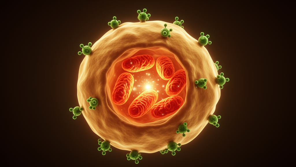
Macrophages are the immune system’s frontline responders, capable of flipping between inflammatory killers and tissue-repairing builders depending on the signals around them. A new study from researchers at the Universidad Nacional Autónoma de México and collaborating institutions shows that a single proinflammatory cytokine, macrophage migration inhibitory factor (MIF), acts less like a command signal that dictates these identities and more like a power supplier that keeps their mitochondria running. In bone marrow-derived macrophages grown in the laboratory, cells lacking MIF still adopted their assigned inflammatory or repair-like profiles, but the inflammatory ones could no longer execute their full program: their reactive oxygen species, mitochondrial superoxide, and nitric oxide output all dropped, alongside weakened mitochondrial membrane potential and impaired oxidative phosphorylation. The work, published in Frontiers in Cell Death, suggests that endogenous MIF safeguards the bioenergetic machinery that inflammatory macrophages need to function at full capacity.
The research team, led by Christian D. Ortiz-Robles and Antonio Andrade-Meza as co-first authors with Miriam Rodríguez-Sosa as corresponding author, cultured macrophages from wild-type BALB/c mice and from mice genetically engineered to lack MIF. They exposed these bone marrow-derived macrophages to four conditions: a nonpolarized basal state called M0, an inflammatory M1 state induced by lipopolysaccharide plus interferon-gamma, a repair-associated M2 state induced by interleukin-4 plus interleukin-13, and a basal state supplemented with recombinant MIF protein. Flow cytometry confirmed that both genotypes differentiated normally under each condition, with M1 markers appearing in roughly 60 to 65 percent of cells under inflammatory stimulation and M2 markers in roughly 28 to 40 percent under repair stimulation, regardless of whether MIF was present. This established a crucial baseline: the absence of MIF did not prevent macrophages from acquiring their assigned surface identities.
What changed was what those cells could actually do. When the researchers measured total intracellular reactive oxygen species using a fluorescent probe, M1-polarized macrophages from MIF-deficient mice produced significantly less than their wild-type counterparts, a partial but meaningful blunting of the oxidative burst that normally accompanies classical activation. Mitochondrial superoxide, measured with a probe targeted specifically to the mitochondrial matrix, followed the same pattern, rising sharply in both genotypes under M1 conditions but reaching significantly lower levels in the MIF-deficient cells. Nitric oxide, quantified in culture supernatants by the Griess method, was strongly induced in both groups under M1 polarization, yet the MIF-deficient macrophages generated significantly less of this antimicrobial and signaling molecule. In contrast, M2-polarized macrophages from both genotypes showed baseline levels of these reactive species, and arginase activity, a hallmark of alternative activation, rose comparably in both wild-type and MIF-deficient M2 cells.
Mitochondrial membrane potential, the electrical charge across the inner mitochondrial membrane that drives ATP synthesis, told a similar story. Using MitoTracker fluorescence and confocal microscopy, the researchers found that M1, M2, and recombinant MIF-treated macrophages lacking MIF all displayed markedly reduced membrane potential compared with their wild-type equivalents. A complementary flow cytometric approach using the JC-10 dye, which shifts from orange-red to green fluorescence when mitochondria depolarize, confirmed these observations, showing a modest but significant increase in depolarized mitochondria across M0, M1, and M2 states in the MIF-deficient cells. Notably, the MIF-deficient macrophages showed both lower reactive oxygen species and lower membrane potential simultaneously, which the authors interpret as evidence of impaired bioenergetic competence rather than oxidative damage causing the depolarization.
To probe respiratory function directly, the team employed Seahorse extracellular flux analysis, measuring oxygen consumption rates in real time while sequentially adding inhibitors that dissect different components of mitochondrial respiration. Wild-type M0 and M2 macrophages maintained robust oxygen consumption between 40 and 60 picomoles of oxygen per minute, while M1 and recombinant MIF-treated cells dropped to 10 to 20 picomoles per minute, reflecting the known metabolic shift of inflammatory macrophages toward glycolysis. When the researchers compared genotypes within each polarization state, basal respiration was significantly lower in MIF-deficient M0, M1, and M2 macrophages than in their wild-type counterparts. ATP-linked respiration, calculated as the drop in oxygen consumption after ATP synthase inhibition, was significantly reduced in MIF-deficient M0 and M2 cells. Maximal respiratory capacity, measured after uncoupling with FCCP, was also lower in MIF-deficient M0 macrophages, indicating that even before any polarization signal arrives, MIF contributes to maintaining the respiratory reserve that mitochondria need.
The ratio of ATP-coupled respiration to maximal respiration provided a window into mitochondrial efficiency. Wild-type M1 macrophages showed a low ratio of 0.19, consistent with their glycolytic bias, while MIF-deficient M1 cells dropped further to 0.16. Wild-type M2 macrophages reached a ratio of 0.52, reflecting their oxidative character, but the MIF-deficient equivalent was consistently lower across all polarization states. Proton leak, the component of respiration that dissipates membrane potential without producing ATP, was markedly reduced in MIF-deficient M1 and M2 macrophages, at 42.8 percent and 58.6 percent of wild-type levels respectively. Because proton leak is partly driven by the magnitude of the membrane potential, these parallel reductions in membrane potential and proton flux reinforce the picture of a mitochondrial system that has lost part of its operating capacity when MIF is absent.
A real-time ATP rate assay separated the contributions of glycolysis and oxidative phosphorylation to total ATP production. Glycolytic ATP was actually higher in MIF-deficient M0 macrophages than in wild-type cells, at 116 percent of the wild-type value, suggesting a compensatory shift toward glucose fermentation when mitochondrial output falls. However, mitochondrial ATP production was reduced to roughly 80 percent of wild-type levels in MIF-deficient M0 cells. Under M2 conditions, where oxidative phosphorylation dominates, the deficit was more pronounced: MIF-deficient M2 macrophages produced only about 44 percent of the mitochondrial ATP generated by their wild-type counterparts. M1 macrophages from both genotypes showed similarly low mitochondrial ATP production, differing by only about 4 percent, which aligns with their reliance on glycolysis rather than oxidative metabolism.
The recombinant MIF experiments added an important nuance. Adding exogenous MIF protein to basal M0 macrophages did not restore mitochondrial function in MIF-deficient cells, nor did it induce reactive oxygen species, superoxide, or nitric oxide production in wild-type cells, even at doses up to 100 nanograms per milliliter. The authors caution that recombinant MIF was added only once at the start of the culture without replenishment, and its stability was not assessed, so the treatment represents a single acute exposure rather than sustained signaling. This limitation means the study cannot determine whether continuous MIF delivery or reintroduction during inflammatory polarization would rescue the observed defects, and the authors explicitly call for future rescue experiments, receptor blockade studies, and time-resolved mitochondrial analysis to address these questions.
The findings position MIF as a permissive metabolic factor rather than a direct polarizing signal. The authors propose that MIF supports the mitochondrial state required for full macrophage effector function, particularly during inflammatory M1 activation, while M2 macrophages can compensate for its absence through metabolic flexibility. They also note that their previous in vivo observations in a Taenia crassiceps infection model align with this interpretation, showing reduced M1 macrophage proportions in MIF-deficient mice during the early inflammatory phase while M2 frequencies remained comparable during chronic infection. Whether the mitochondrial defects observed in cultured bone marrow-derived macrophages also occur in tissue-resident or infection-induced macrophages within living animals remains an open question, as does the precise mechanism by which MIF influences mitochondrial function, with CD74-dependent signaling and p53 regulation among the candidate pathways the authors suggest warrant direct experimental testing.
Subject of Research: The role of macrophage migration inhibitory factor in supporting mitochondrial metabolism during inflammatory macrophage activation
Article Title: Macrophage migration inhibitory factor (MIF) supports mitochondrial metabolism during inflammatory macrophage activation
Article References: Ortiz-Robles, C. D., Andrade-Meza, A., Rodríguez, T., Ortiz-Sánchez, B. J., Juárez-Avelar, I., Gutiérrez-Cirlos, E. B., Maycotte, P., Calderón-Aranda, E. S., & Rodríguez-Sosa, M. (2026). Macrophage migration inhibitory factor (MIF) supports mitochondrial metabolism during inflammatory macrophage activation. Frontiers in Cell Death, 5, 1892088. https://doi.org/10.3389/fceld.2026.1892088
Image Credits: AI Generated
DOI: 10.3389/fceld.2026.1892088
Keywords: macrophage migration inhibitory factor, macrophage polarization, mitochondrial metabolism, oxidative phosphorylation, reactive oxygen species, nitric oxide, M1 macrophages, M2 macrophages, bioenergetics, membrane potential, ATP production, inflammation
News Source: Drew Townsend. (October 30, 2026). Immune Cytokine MIF Keeps Macrophage Mitochondria Firing During Inflammation. Scienmag.



