


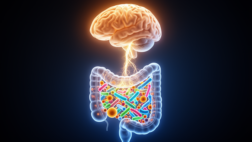
The trillions of bacteria living in the human gut are emerging as unexpected players in some of the most feared diseases of the brain. A new review published in Current Genetics on 21 August 2025 pulls together the evidence connecting the human microbiome to Alzheimer’s disease, Parkinson’s disease, Huntington’s disease and amyotrophic lateral sclerosis, and argues that the microbial communities colonising the body since birth may shape how these neurodegenerative conditions begin and progress. Written by Nilabha Mukherjea, Ashwin Khandelwal, Rohit Saluja and Neetu Kalra, the perspective article stresses that the field is still young, with many findings preliminary and inconsistent, but the accumulating data point to a genuine biological connection worth serious attention.
The scale of the microbial world inside us is staggering. The human body contains roughly 30 trillion human cells alongside about 38 trillion bacterial cells, and the gastrointestinal tract alone harbours more than 200,000 diverse, non-redundant prokaryotic genomes spanning around 5,000 different microbial species, which together encode nearly 200 million distinct protein sequences. The gut microbiome is dominated by four bacterial phyla: Firmicutes, Bacteroidetes, Actinobacteria and Proteobacteria. These communities are not static. An infant’s first microbiota depends heavily on the mode of delivery, with vaginally born babies acquiring microbes resembling their mothers’ vaginal flora and caesarean-delivered infants inheriting communities closer to skin microbes. Diversity grows through childhood with breast milk, solid foods, antibiotic exposure and eventually an adult diet, and it shifts again in later life, when the balance is disturbed and Proteobacteria levels rise.
Why should gut bacteria matter for the brain? The answer lies in the gut–brain axis, a bidirectional communication pathway linking the gastrointestinal tract, the microbiome and the central nervous system through the enteric nervous system, the vagus nerve, the immune system and the circulation. Microbial metabolites, gut hormones and neuroactive substances travel along these routes and influence brain function. The brain, in turn, regulates gut activity through the hypothalamic–pituitary–adrenal axis and the autonomic nervous system; the neurotransmitter norepinephrine, released during stress, has even been found to promote the proliferation of gut pathogens. Microbial metabolites such as short-chain fatty acids can cross the blood–brain barrier and interact with microglia, the brain’s resident immune cells, while gut bacteria also influence tryptophan metabolism, which underpins the synthesis of serotonin, a neurotransmitter central to mood regulation and cognition.
When this finely tuned system falls out of balance, a state researchers call dysbiosis, the consequences can ripple outward. Dysbiosis is associated with a leaky gut, in which faulty tight junctions increase intestinal permeability, and with the production of harmful metabolites that activate immune cells and drive chronic inflammation. Harmful microbial products released from bacterial cell walls, including lipopolysaccharides, enter systemic circulation and are thought to promote inflammation and release neurotoxins that contribute to neurological disorders. Dysbiosis can also compromise the blood–brain barrier itself: studies in germ-free and antibiotic-treated mice have shown impaired barrier function, supporting the idea that a healthy gut microbiome helps maintain the barrier’s integrity. The review’s authors are careful to note, however, that these results remain largely speculative in humans, and that confirming microbial effects in clinical studies is a significant challenge.
Alzheimer’s disease offers some of the most concrete human evidence. The neuropathology of Alzheimer’s is defined by the aggregation of amyloid-β, followed by hyperphosphorylated tau protein, plaques and neurofibrillary tangles that drive synapse loss and neuronal death. In one human study, patients with amyloid deposits in their brains had decreased levels of Eubacterium rectale and Bacillus subtilis and elevated levels of Escherichia/Shigella in their faeces compared with controls, and the trial also pointed to amyloid and bacterial concentrations contributing to cognitive impairment. Elderly patients with Alzheimer’s tend to have a low proportion of butyrate-producing bacteria, a shortfall that may promote a pro-inflammatory state in both gut and brain, and they show a disrupted Firmicutes to Bacteroidetes ratio, with reduced Firmicutes and increased Bacteroidetes. Metabolic molecules originating from microbes have been reported to associate with biomarkers of phosphorylated tau, amyloid-β load and activation of the NLRP3 inflammasome pathway.
Parkinson’s disease shows perhaps the most striking gut connection of all. Deposits of α-synuclein, the neuropathological hallmark of Parkinson’s, were found in the submucosal and myenteric plexus of the gut before their presence was detected in the brain, hinting that disease activity may spread from gut to brain. Around 80 percent of individuals with Parkinson’s experience prolonged constipation before diagnosis. Human sequencing studies have revealed consistent microbial shifts: patients show reduced levels of Faecalibacterium, Prevotella, Lachnospiraceae, Blautia and Roseburia, bacteria responsible for producing short-chain fatty acids, alongside increases in Lactobacillus and Akkermansia. One study sequencing 72 human Parkinson’s microbiota found a high count of Enterobacteriaceae in patients’ faeces, correlating with postural instability and walking problems, and a 77.6 percent decrease in Prevotellaceae abundance compared with healthy controls. Reduced short-chain fatty acid production is linked to increased gut permeability, which allows toxins and pathogens to enter the bloodstream, initiating neuroinflammation and potentially contributing to the spread of α-synuclein aggregates.
Huntington’s disease and ALS have received far less microbiome scrutiny, but early findings are intriguing. Huntington’s is an autosomal-dominant disorder caused by expanded CAG repeats in the huntingtin gene, producing polyglutamine-expanded nonfunctional huntingtin protein that forms aggregates and damages neurons. The first study linking Huntington’s to the gut microbiome found that patients showed low alpha diversity and higher beta diversity than controls, along with a major shift in overall microbial community structure at the phylum and family level. In a mouse model, control animals showed high diversity and abundance in Firmicutes whereas Huntington’s mice showed low overall diversity and abundance in Bacteroidetes. In ALS, a progressive loss of motor neurons, growing evidence indicates that gastrointestinal symptoms precede neurological symptoms. An ALS mouse model showed a significant decrease in butyrate-producing bacteria such as Butyrivibrio, which affected intestinal tight junctions and led to leaky gut and inflammation. In antibiotic-treated ALS mice, Akkermansia muciniphila ameliorated symptoms by triggering accumulation of nicotinamide in the central nervous system, whereas Ruminococcus torques and Parabacteroides distasonis worsened them. Human ALS patients’ faecal samples showed less diverse gut bacteria than those of healthy individuals, with a notably low Firmicutes to Bacteroidetes ratio.
The review is refreshingly candid about how messy the evidence remains. The studies by Cattaneo and colleagues and by Zhan and colleagues involved limited numbers of patients and lacked reproducibility, and the bacterial taxa associated with plaque formation were not consistent across studies. In Parkinson’s research, findings are similarly inconsistent: one study reported a microbiome composition markedly different from others, and a study of Egyptian patients found no significant differences from controls except a high Prevotella to Bacteroides ratio. A metagenomic analysis examining associations between ten diseases and the microbiome found that disease-associated differences are not consistent across studies, reflecting variability across cohorts, methodologies and sequencing platforms. Animal models add another layer of difficulty, because many lack bacteria found in humans; Bacteroides species, for example, are dominant in humans but rare in laboratory mice, and symptoms such as mood changes and cognitive impairment cannot be measured in mice with the detail possible in people.
Therapeutic possibilities are nonetheless advancing. Probiotics and nutrient-rich diets have been reported to promote Bifidobacteria, reduce inflammation and lower amyloid-β deposition in Alzheimer’s, leading to cognitive improvements. In Parkinson’s, formulations containing Lactobacillus acidophilus and Bifidobacterium infantis significantly decreased pain and abdominal bloating in patients, a randomised trial with multi-strain probiotics significantly improved stool consistency and frequency, and an innovative diagnostic approach based on Prevotellaceae levels and constipation status yielded a specificity rate of 90.3 percent. Fecal microbiota transplantation in a Parkinson’s patient showed improvement in motor functions and reduction in inflammation. In Huntington’s, a six-week probiotic capsule trial showed no significant changes in patients, which the study attributed to the limited treatment duration. In ALS, animal and early human trials suggest fecal microbiota transplantation and probiotics may improve motor function and reduce inflammation via immune modulation, and an ongoing clinical trial is testing fecal microbiota transplantation in ALS patients.
The authors’ conclusion is measured rather than triumphant. The central question in human microbiota research, they write, is whether the gut microbiome is enough to allow effective clinical interventions that improve patient outcomes. Translating microbiome-based therapies into practice is challenged by key differences between animal and human models, including site-specific fermentation and variations in microbial composition and intestinal cell types. They call for multi-omics integration and standardised bioinformatics to unravel microbiome functionality and ensure reproducible results, alongside longitudinal studies and organ-on-a-chip technologies to enhance translational accuracy. The lifelong variability and individual specificity of the gut microbiome, shaped by factors like diet and geography, supports personalised therapeutic approaches, but caution is vital during preclinical development to monitor and minimise potential side effects of microbiome-targeted therapies. What is clear is that the role of the human microbiome in neurodegenerative disease is genuine, even if the mechanisms remain to be fully understood, and every step in charting the fate of the microbiota and its metabolites brings researchers closer to better diagnosis and treatment.
Subject of Research: The role of the human gut microbiome in neurodegenerative diseases
Article Title: The Role of the human microbiome in neurodegenerative diseases: A Perspective
Article References: Mukherjea, N., Khandelwal, A., Saluja, R., & Kalra, N. (2025). The Role of the human microbiome in neurodegenerative diseases: A Perspective. Current Genetics, 71(1), Article 17. https://doi.org/10.1007/s00294-025-01319-8
Image Credits: AI Generated
DOI: 10.1007/s00294-025-01319-8
Keywords: human microbiome, gut–brain axis, Alzheimer's disease, Parkinson's disease, Huntington's disease, amyotrophic lateral sclerosis, dysbiosis, short-chain fatty acids, neuroinflammation, probiotics, fecal microbiota transplantation, blood–brain barrier
News Source: Cassandra Pierce. (October 30, 2026). Gut microbes linked to Alzheimer’s, Parkinson’s, Huntington’s and ALS. Scienmag.



