Repeated peripheral exposure to histamine left a lasting imprint on the brain’s innate immune cells in mice and was associated with a milder response to a subsequent dopaminergic injury, according to an open-access original research article published on 4 November 2026 in the journal Cellular and Molecular Neurobiology. The study, authored by Ana Margarida Vale, Catarina Serra-Almeida, Jorge Valero, Liliana Bernardino and colleagues, examined whether inflammatory priming of the periphery could reshape glial cells in defined brain regions and alter how the nigrostriatal system copes with a later insult.
The researchers built their experiment around a concept already explored in other neurological conditions. Peripheral immune priming can induce long-lasting changes in brain innate immune responses, and in diseases including Alzheimer’s disease and stroke, inflammatory priming has been associated with neuroprotection, likely through microglial reprogramming. The team set out to test whether repeated peripheral histamine exposure would be associated with persistent glial morphometric remodelling and with an altered response to subsequent dopaminergic injury in a Parkinson’s disease model.
Adult male mice received four consecutive daily intraperitoneal injections of histamine, lipopolysaccharide, or PBS as a control. Lipopolysaccharide, commonly abbreviated LPS, served as a reference peripheral immune-stimulating condition. Three weeks after this priming phase, the investigators assessed glial morphometry in two regions of the nigrostriatal pathway: the substantia nigra, which houses the dopamine-producing neurons lost in Parkinson’s disease, and the striatum, their principal target area.
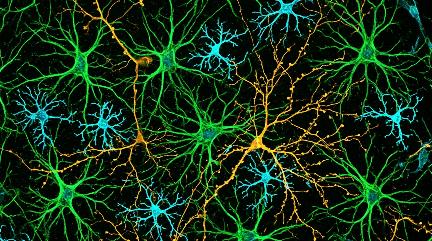
The morphometric results were strikingly region-specific. Under non-lesioned conditions, both histamine and LPS exposure were associated with persistent microglial morphometric remodelling in the substantia nigra. In the striatum, however, comparable microglial changes were detected only after LPS, not after histamine. Astrocytic measures based on glial fibrillary acidic protein, or GFAP, showed only modest, non-significant trends in the substantia nigra, indicating that the clearest priming effects at this stage involved microglia rather than astrocytes.
To probe whether this remodelling mattered functionally, the team used a separate cohort of mice. These animals underwent an intrastriatal 6-hydroxydopamine lesion three weeks after priming. 6-hydroxydopamine, or 6-OHDA, is a toxin that selectively destroys catecholaminergic neurons and is widely used to model the dopaminergic degeneration of Parkinson’s disease in rodents. Two weeks after the lesion, the researchers evaluated motor behaviour, nigrostriatal tyrosine hydroxylase immunoreactivity, apomorphine-evoked striatal neuronal recruitment and the glial morphometric response.
Behavioural and histological outcomes pointed in the same direction. Following the 6-OHDA lesion, prior exposure to either histamine or LPS reduced apomorphine-induced rotational asymmetry, a standard motor readout in lesioned mice, and was associated with greater preservation of tyrosine hydroxylase immunoreactivity in the nigrostriatal system. Tyrosine hydroxylase, or TH, is the rate-limiting enzyme in dopamine synthesis, so its immunoreactivity serves as a proxy for the integrity of dopaminergic fibres and neurons.
Histamine priming produced additional effects beyond those shared with LPS. Mice primed with histamine showed increased apomorphine-evoked recruitment of DARPP-32-positive striatal neurons, a measure of striatal neuronal responsiveness to dopamine receptor stimulation. Histamine priming also attenuated the lesion-associated microglial changes in both the substantia nigra and the striatum. The astrocytic responses after the lesion were region-dependent, differing between the two examined areas rather than following a single uniform pattern.
Taken together, the findings indicate that repeated peripheral histamine exposure was associated with persistent, region-specific glial morphometric remodelling and with attenuated behavioural and dopaminergic deficits following subsequent 6-OHDA injury. The authors describe these results as compatible with long-lasting histamine-associated neuroimmune conditioning, a state in which peripheral immune stimulation appears to reconfigure how resident glial cells respond to a later challenge.
The study’s authors are careful about what these associations do and do not establish. The underlying cellular and molecular mechanisms remain to be determined, and the causal contribution of the observed glial remodelling to the protective pattern has not yet been demonstrated. The work was conducted exclusively in adult male mice with a toxin-based lesion model, so the findings are preclinical observations rather than evidence about therapeutic effects in humans. Because the article was shared early as peer-reviewed, accepted research carrying a permanent DOI, with a final version of record to follow, readers should also note that the text remains subject to further edits.
Even with those caveats, the report adds to a growing body of work suggesting that signals originating outside the brain can leave durable marks on its resident immune cells. If the mechanisms behind histamine-associated conditioning can be identified, the region-specific nature of the remodelling described here may offer clues about why some glial responses correlate with preserved dopaminergic circuitry while others do not, a question the authors flag as central to future work.
Subject of Research: Peripheral histamine priming, glial remodelling and dopaminergic injury in a mouse model of Parkinson's disease
Article Title: Histamine-Induced Peripheral Priming Promotes Region-Specific Glial Remodelling and Protects Against Dopaminergic Injury in a Mouse Model of Parkinson’s Disease
Article References: Vale, A. M., Serra-Almeida, C., Valero, J., & Bernardino, L. (2026). Histamine-Induced Peripheral Priming Promotes Region-Specific Glial Remodelling and Protects Against Dopaminergic Injury in a Mouse Model of Parkinson’s Disease. Cellular and Molecular Neurobiology. https://doi.org/10.1007/s10571-026-01845-0
Image Credits: AI Generated
DOI: 10.1007/s10571-026-01845-0
Keywords: histamine, peripheral immune priming, microglia, astrocytes, substantia nigra, striatum, 6-hydroxydopamine, Parkinson's disease, neuroinflammation, dopaminergic neurons, mouse model, glial morphometry
News Source: Cassandra Pierce. (November 5, 2026). Repeated Histamine Exposure Shows Lasting Glial Changes and Attenuated Dopaminergic Deficits in Mice. Scienmag.