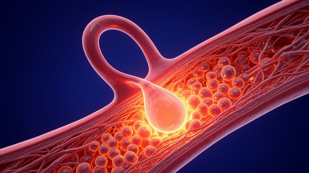
A systematic literature review published in Artery Research synthesizes fifteen years of cerebrovascular investigations, revealing that intracranial aneurysms develop and rupture through the coordinated execution of multiple programmed cell death pathways. Characterized by localized abnormal bulging of intracranial arterial walls, these vascular lesions pose severe clinical consequences, primarily when rupture causes catastrophic events such as subarachnoid hemorrhage and intracerebral hematoma. In clinical observations, subarachnoid hemorrhage occurs in as many as ninety percent of ruptured middle cerebral artery aneurysms, which represent a major subtype accounting for twenty to forty percent of all intracranial aneurysm presentations. Acute-phase mortality following middle cerebral artery aneurysm rupture ranges between thirty and fifty percent, while forty to sixty percent of surviving individuals experience permanent neurological deficits, including limb hemiplegia, cognitive impairment, and aphasia. The synthesized evidence demonstrates that aneurysm pathogenesis extends beyond passive wall weakening, acting instead as an active, highly regulated biological destruction of the arterial wall driven by apoptosis, ferroptosis, pyroptosis, and autophagy-dependent cell death.
Anatomical analyses emphasize that middle cerebral artery aneurysms display pronounced site specificity, with more than eighty percent of lesions arising at the bifurcation zone where the main M1 segment branches into the M2 segment. Arterial bifurcations around the circle of Willis inherently harbor developmental vulnerabilities, such as discontinuous internal elastic lamina and sparse medial smooth muscle cell distribution. When blood flows from the internal carotid artery, the diversion generates intense wall shear stress and oscillatory hemodynamic disturbance across this bifurcation, superimposing mechanical stress upon preexisting congenital gaps. Clinical neuroimaging confirms that while most lesions present as saccular protrusions, certain middle cerebral artery aneurysms exhibit a lobulated morphology, which demonstrates an annual rupture rate 2.3 times higher than that of ordinary saccular aneurysms. Regional variations further influence detection, with middle cerebral artery aneurysms accounting for twenty-two to 31.5 percent of intracranial aneurysms globally and reaching up to forty-five percent in specific Asian cohorts.
Current neurosurgical practice remains constrained by a recognized triple management dilemma consisting of passive observation during asymptomatic phases, substantial procedural hazards following rupture, and an absence of precise molecular targets. Asymptomatic intracranial aneurysms are predominantly detected incidentally during unrelated imaging evaluations, leaving patients dependent on serial computed tomography angiography, magnetic resonance angiography, or digital subtraction angiography every six to twelve months. This surveillance approach cannot eliminate sudden rupture events, as asymptomatic middle cerebral artery aneurysms exceeding seven millimeters in diameter maintain an annual rupture rate between 1.5 and 2.0 percent. When intervention becomes necessary, microsurgical craniotomy clipping carries a postoperative neurological impairment rate of twenty-five to thirty percent for deep lesions adjacent to critical motor or language areas. Alternatively, endovascular interventional embolization presents a postoperative recurrence rate of ten to fifteen percent in wide-necked or irregularly shaped aneurysms, accompanied by considerable procedural expenses.
To evaluate the biological drivers underpinning these clinical challenges, the review authors executed a systematic literature screening protocol covering studies indexed between January 2010 and December 2025 across PubMed, Web of Science, and Embase. Search architectures paired core descriptors, including intracranial aneurysm, middle cerebral artery aneurysm, and programmed cell death, with secondary terms covering apoptosis, ferroptosis, pyroptosis, autophagy, pathogenesis, and vascular wall cells. The investigators conducted a structured two-stage screening process: the primary author executed preliminary assessments of titles and abstracts to exclude duplicate publications, small-sample case reports with fewer than ten cases, non-systematic reviews, and articles outside Chinese or English languages, followed by full-text methodological evaluations performed with invited co-authors to verify experimental validity, design integrity, and objective relevance.
Histopathological and molecular examinations demonstrate that arterial wall weakening involves coordinated structural degeneration across the intima, media, and adventitia, primarily driven by cellular apoptosis. Pathological tissue specimens reveal fragmented elastic lamina, culminating in complete segmental absence of internal elastic lamina at the neck of ruptured middle cerebral artery aneurysms alongside heavy local concentrations of apoptotic cells. Within the tunica media, apoptosis causes selective depletion of vascular smooth muscle cells, reducing smooth muscle cellularity by more than fifty percent relative to healthy arterial tissue. Because vascular smooth muscle cells provide the primary contractile support and mechanical tension required to withstand intraluminal pressure, this cellular loss weakens the arterial architecture, replacing dense muscular layers with disorganized collagen matrices. Concurrently, high wall shear stress, oxidative stress, and inflammatory mediators trigger apoptosis and structural detachment of luminal endothelial cells, downregulating tight junction proteins including occludin and claudin, which breaches the protective vascular barrier and fuels further cellular injury.
Within the aneurysm microenvironment, endothelial cells and vascular smooth muscle cells actively orchestrate vascular remodeling through dynamic signaling cascades. Upon experiencing abnormal shear stress, endothelial cells activate nuclear factor kappa B signaling via membrane integrins and cytoskeletal remodeling, upregulating vascular cell adhesion molecule-1 and intercellular adhesion molecule-1 to recruit circulating leukocytes and macrophages. Endothelial cells simultaneously generate reactive oxygen species that diminish nitric oxide bioavailability, compromise vasodilation, and initiate endothelial-to-mesenchymal transition, wherein endothelial cells lose specific markers like CD31 and VE-cadherin while acquiring mesenchymal properties marked by alpha-smooth muscle actin and fibronectin. In response to inflammatory cytokines such as interleukin-1 beta, tumor necrosis factor-alpha, and platelet-derived growth factor, medial smooth muscle cells undergo phenotypic switching from a contractile state into a synthetic state. Although synthetic smooth muscle cells produce extracellular matrix components, an overexpression of matrix metalloproteinases MMP-2 and MMP-9 against inadequate tissue inhibitors of metalloproteinases accelerates structural matrix breakdown, which adventitial fibroblast and pericyte apoptosis further compounds.
Mitochondria-dependent intrinsic apoptosis serves as a primary executioner of vascular wall degradation, triggered by local tissue ischemia, hypoxia, hemodynamic strain, and inflammatory signaling. Luminal stenosis and local wall thickening compress the vasa vasorum in middle cerebral artery aneurysm lesions, starving intramural cells of oxygen and elevating hypoxia-inducible factor-1 alpha, which directly stimulates pro-apoptotic Bax transcription while suppressing anti-apoptotic Bcl-xL. Oxidative stress stimulates p53 binding to the Bax promoter, while c-Jun N-terminal kinase activation phosphorylates Bcl-2, neutralizing its anti-apoptotic activity. Consequently, Bax and Bak oligomerize across the outer mitochondrial membrane, inducing outer membrane permeabilization and discharging cytochrome c, Smac/DIABLO, and apoptosis-inducing factor into the cytosol. Cytosolic cytochrome c binds apoptotic protease-activating factor-1 and adenosine triphosphate to assemble the apoptosome, initiating Caspase-9 cleavage and activating downstream Caspase-3, with clinical middle cerebral artery aneurysm tissue demonstrating Caspase-9 and Caspase-3 enzymatic activity four to six times higher than normal intracranial arterial tissue.
Beyond classical apoptosis, ferroptosis significantly accelerates structural arterial wall compromise through iron-dependent lipid peroxidation and metabolic imbalance. Following initial endothelial injury, the degradation of extravasated hemoglobin liberates free iron, which enters cells via transferrin receptor 1 while impaired ferritin heavy and light chains fail to sequester iron surpluses, catalyzing hydroxyl radical generation through Fenton chemistry. Concurrently, inflammatory mediators suppress the System Xc- cystine-glutamate antiporter subunits SLC7A11 and SLC3A2, depleting intracellular glutathione stores and inactivating glutathione peroxidase 4, the primary enzymatic defender against lipid membrane oxidation. Polyunsaturated fatty acids in vascular cell membranes are subsequently processed by acyl-CoA synthetase long-chain family member 4, driving extensive lipid ROS accumulation and triggering membrane disintegration. Bioinformatics analyses confirm that competing endogenous RNA regulatory networks, including PVT1-miR-4644-SLC39A14 and DUXAP8-miR-378e/378f-SLC2A3, accelerate iron overload in middle cerebral artery aneurysms, where clinical samples exhibit elevated iron deposits and gene expression patterns that correlate positively with rupture risk.
Pyroptosis acts as a pro-inflammatory cell death modality that amplifies vascular wall remodeling through inflammasome assembly and cytokine processing. In damaged aneurysm walls, pattern recognition receptors like Toll-like receptor 4 recognize damage-associated molecular patterns, activating nuclear factor kappa B to prime transcription of NLRP3, ASC, and pro-Caspase-1. Subsequent extracellular adenosine triphosphate binding to purinergic P2X7 receptors induces potassium efflux, triggering NLRP3 oligomerization with ASC and pro-Caspase-1 into functional inflammasomes. Assembled Caspase-1 undergoes autocatalytic cleavage to yield active p20/p10 heterodimers, which cleave Gasdermin D to release its N-terminal fragment. These fragments insert into cell membranes to form pores measuring ten to twenty nanometers in diameter, causing cell swelling, osmotic lysis, and the release of mature interleukin-1 beta and interleukin-18. The released interleukin-1 beta subsequently upregulates matrix metalloproteinase-9 expression, accelerating elastin and collagen degradation, while estrogen-deficient models reveal that downregulation of estrogen receptor alpha and Sirt1 further amplifies NLRP3/IL-1beta/MMP-9 signaling.
Autophagy exhibits a stage-dependent, bidirectional influence on intracranial aneurysm integrity, transitioning from early cellular protection to late-stage structural destruction. During initial disease stages, basal autophagy preserves cellular homeostasis by clearing damaged organelles and selectively degrading the transcription factor FOXO3. As chronic inflammation and oxidative stress intensify, sustained autophagic inhibition leads to pathological FOXO3 accumulation, which transcriptionally upregulates the pro-apoptotic gene BBC3/PUMA and heightens smooth muscle cell vulnerability to apoptotic death. Simultaneously, oxidative stress stimulates the PINK1/Parkin signaling cascade to drive excessive mitophagy, while FAM134B upregulation mediates endoplasmic reticulum autophagy, culminating in mitochondrial energy collapse and intracellular release of cytochrome c and mitochondrial DNA. Clinical tissue analyses identify marked elevations of autophagy-dependent cell death markers including Atg5, Beclin1, FOXO3, and PUMA in middle cerebral artery aneurysm walls, correlating directly with increased rupture probability.
Experimental preclinical studies have explored targeted interventions to counteract these cell death cascades and restore cellular equilibrium. Adeno-associated virus-mediated Bcl-2 overexpression in animal models reduces smooth muscle cell apoptosis by more than forty percent, an effect that increases by an additional eight to ten percent in middle cerebral artery aneurysms, curbing aneurysm expansion by thirty-five percent. Intraperitoneal delivery of the broad-spectrum Caspase inhibitor Z-VAD-FMK penetrates the blood-brain barrier to reduce intracranial aneurysm rupture rates by twenty-eight percent in rats, reaching a thirty-two percent reduction in middle cerebral artery lesions. Upstream modulation via the Akt agonist SC79 lowers smooth muscle cell apoptotic indices by forty-two to forty-five percent while reducing lesion progression by fifty percent, whereas exosomal delivery of miR-126 suppresses Caspase-3 to enhance endothelial barrier integrity by thirty to thirty-five percent. Furthermore, repurposing conventional pharmacological agents shows documented efficacy: retrospective clinical analyses demonstrate that long-term atorvastatin therapy correlates with a thirty-two percent slower aneurysm progression rate and a twenty percent lower rupture risk overall, expanding to thirty-six percent and twenty-four percent respectively in middle cerebral artery aneurysms, while low-dose aspirin reduces mural inflammation by forty percent in animal models.
Despite promising preclinical findings, the review underscores substantial translational obstacles that currently prevent clinical implementation. Available interventions remain restricted to cellular cultures and small-animal models, such as surgically induced rat aneurysms, which experience acute, unifactorial development that fails to replicate the chronic, multi-decade progression of human vascular lesions. Furthermore, non-specific pharmacological agents carry notable toxicological risks; broad-spectrum Caspase inhibition disrupts physiological cell clearance and elevates oncogenic hazards, while excessive PI3K/Akt activation risks inducing pathological vascular stenosis. Future translational progress depends on integrating single-cell RNA sequencing and spatial transcriptomics to resolve cell-specific targets, such as GPX4 downregulation in smooth muscle cells and Fas upregulation in endothelial cells. Developing intelligent, microenvironment-responsive nanocarriers modified with VEGFR2 or PDGFR-beta ligands, establishing standardized miniature pig models, and validating circulating biomarkers will be critical to transforming programmed cell death research into individualized, non-invasive therapies for high-risk intracranial aneurysms.
Subject of Research: Programmed cell death mechanisms and vascular wall degradation in intracranial aneurysms
Article Title: Programmed Cell Death: A Key Pathway in Intracranial Aneurysm Progression
Article References: Li, Y., Zeng, Q., Ding, D., Zheng, K., Xiong, J., Sun, J., & Tong, X. (2026). Programmed Cell Death: A Key Pathway in Intracranial Aneurysm Progression. Artery Research, 32(1), Article 26. https://doi.org/10.1007/s44200-026-00119-w
Image Credits: AI Generated
DOI: 10.1007/s44200-026-00119-w
Keywords: Intracranial aneurysms, Middle cerebral artery aneurysms, Programmed cell death, Apoptosis, Ferroptosis, Pyroptosis, Autophagy-dependent cell death, Vascular smooth muscle cells, Vascular endothelial cells, Subarachnoid hemorrhage, Internal elastic lamina, NLRP3 inflammasome, Gasdermin D, Glutathione peroxidase 4, Reactive oxygen species, Bcl-2 family proteins, Matrix metalloproteinases, Caspase cascades
News Source: Ophelia Keating. (November 16, 2026). Programmed Cell Death Pathways Drive Intracranial Aneurysm Progression and Rupture Risk. Scienmag.