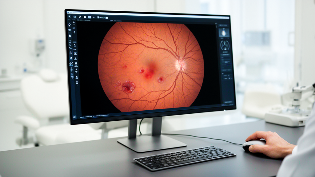
A recent systematic review examining seventy-eight research papers has evaluated the current landscape of machine learning and deep learning applications in the screening, grading, and lesion segmentation of diabetic retinopathy. The study, which adheres to Preferred Reporting Items for Systematic Reviews and Meta-Analyses guidelines, characterizes the efficacy of various computational models while highlighting significant challenges related to data quality, model complexity, and the translation of automated methods into practical clinical environments. Diabetic retinopathy remains a leading global cause of blindness, necessitating early identification and prompt treatment to prevent vision impairment. The manual screening of retinal fundus images is inherently time-consuming and tedious, creating a profound diagnostic gap between the millions of patients affected by diabetes and the limited number of available medical experts, particularly in low-income regions where the ratio of ophthalmologists to patients remains significantly skewed. This scarcity of clinical resources underscores the potential utility of artificial intelligence systems to facilitate precise, efficient, and cost-effective automated screening.
The researchers conducted a comprehensive literature search across major databases including PubMed, IEEE Xplore, ScienceDirect, and Scopus to isolate pertinent studies that applied machine learning or deep learning to the detection, classification, and segmentation of diabetic retinopathy. By applying rigorous inclusion and exclusion criteria, the team narrowed the focus to seventy-eight distinct research papers that provided sufficient evidence on performance metrics, modeling methodologies, and image preprocessing techniques. The evaluation focused on key retinal datasets such as APTOS 2019, IDRiD, MESSIDOR, and EyePACS, which serve as foundational resources for training neural networks. These datasets vary significantly in scale, image resolution, and clinical categorization, highlighting the variability in quality that impacts the development of robust, generalizable diagnostic algorithms. The study observed that while datasets like APTOS 2019 are frequently utilized for binary classification, others provide finer annotations for lesion-specific segmentation, each presenting unique challenges for model training.
Effective image preprocessing remains a critical stage in the automated analysis of retinal fundus images, as raw diagnostic data is often degraded by artifacts such as environmental factors, camera lens dust, eye blinking, or improper flash settings. The review identifies common intensity transformation techniques, including Contrast Limited Adaptive Histogram Equalization, which is widely employed to enhance local contrast without amplifying existing noise. These preprocessing steps directly influence model accuracy, as demonstrated by studies that applied similar convolutional neural network architectures to the same dataset but yielded divergent performance outcomes depending on the chosen enhancement protocols.
Binary classification studies aim to categorize images as either healthy or indicating the presence of diabetic retinopathy. Binary classification models often leverage handcrafted features, such as local binary patterns and histograms of oriented gradients, combined with support vector machines to achieve competitive performance, particularly when computational resources are limited. However, the field has increasingly shifted toward deep learning architectures, such as EfficientNet, ResNet, and Inception-V3. While these pre-trained models demonstrate high diagnostic accuracy, they often carry a substantial number of trainable parameters. This high parameter count introduces significant memory footprint and latency constraints, raising concerns regarding their deployment on resource-limited devices. Recent research has attempted to mitigate these issues by proposing lightweight custom-designed convolutional neural networks that utilize skip connections to maintain gradient flow during backpropagation, thereby reducing the overall model size without sacrificing diagnostic efficacy.
The objective of diabetic retinopathy grading is to categorize the severity of the disease, often using systems like the International Clinical Diabetic Retinopathy classification, which ranges from mild non-proliferative retinopathy to proliferative forms. Automated grading approaches have evolved from custom convolutional neural networks to complex ensemble methods that integrate diverse architectures, such as VGG, DenseNet, and Inception models. Attention mechanisms, including residual, spatial, and channel attention, are now standard in state-of-the-art grading frameworks to help models focus on tiny lesions like microaneurysms, which might otherwise be overlooked. Some researchers have integrated classical machine learning classifiers like extreme learning machines or J48 algorithms at the output stage, demonstrating that hybrid approaches can provide high performance while potentially reducing the complexity associated with pure deep learning models.
Segmentation is essential for isolating regions of interest, including the optic disc, fovea, and specific pathological lesions such as hemorrhages, hard exudates, and microaneurysms. The UNet model, known for its encoding-decoding path and skip connections, serves as a popular foundation for these tasks, with variants such as UNet++ and dense residual path UNet achieving high performance in blood vessel segmentation. Lesion-based segmentation specifically requires models capable of detecting small features that exhibit high intra-class variation. Advanced frameworks like Deformable Ladder Bi-attention U-Net have been introduced to address these complexities, utilizing deformable convolutions to capture lesion variations and bi-attention mechanisms to refine contour reconstruction. Despite these advancements, lesion-based segmentation performance remains highly dependent on the quality of the dataset, with some studies reporting variations in Dice scores ranging from 0.579 to 0.998 for different lesions, highlighting the persistent challenge posed by structures that are visually similar to retinal noise.
Recent AI trends in the field are shifting toward addressing the opaque, black-box nature of deep learning models and the constraints of centralized training. Explainable artificial intelligence techniques, including Gradient-weighted Class Activation Mapping and Shapley Additive Explanations, are gaining traction as essential tools for providing visual insights into a model’s decision-making process. By highlighting the specific regions of interest that contribute to a diagnosis, these methods help clinicians understand and trust model outputs, a necessary precursor for deployment in sensitive clinical domains. Simultaneously, federated learning has emerged as a decentralized approach to model training, allowing multiple healthcare institutions to collaboratively develop diagnostic algorithms without sharing raw, sensitive patient data. This strategy not only preserves privacy but also enhances the generalizability of models across diverse populations and clinical conditions, offering a scalable path forward for resource-constrained environments.
The computational complexity of the reviewed models serves as a primary metric for determining their suitability for practical applications, particularly for edge computing and real-time clinical monitoring. The review highlights that while the number of trainable parameters is a consistent and accessible indicator of resource intensiveness, most studies do not explicitly report on other crucial variables such as memory consumption or inference time. This limitation makes it difficult to conduct a standardized comparative analysis of the real-world efficiency of proposed models. Consequently, there is an increasing demand for knowledge distillation, model pruning, and sparsification techniques that can shrink large models without significantly degrading their sensitivity or specificity.
A critical gap identified by the systematic review is the lack of clinical validation for the majority of machine learning models described in the literature. While many algorithms demonstrate excellent performance on benchmark datasets, their efficacy in real-world clinical settings, where imaging variability and anatomical pathologies are more pronounced, remains largely unproven. The review underscores that automated screening systems must be evaluated in prospective clinical studies and developed in close collaboration with ophthalmologists to ensure they address real clinical workflows rather than serving as isolated academic experiments. The reliance on publicly available datasets, which frequently suffer from class imbalance and inconsistent grading standards, limits the ability to measure the true robustness of current models. Researchers are urged to prioritize the development of well-curated, high-resolution datasets that better mirror the complexities of actual diagnostic environments.
Future directions for research must prioritize the creation of lightweight models that can function effectively on edge devices while incorporating federated learning and explainable artificial intelligence to overcome barriers to clinical adoption. By focusing on model interpretability and cross-institutional collaboration, the field can build trust with healthcare providers and move toward integrated diagnostic systems that provide reliable, actionable insights for patient care. Moving forward, the involvement of clinical experts in defining use cases and validating performance in practical settings remains essential for achieving true ophthalmologist-level diagnostic grading that can be reliably deployed at scale.
Subject of Research: Systematic review of machine learning applications for diabetic retinopathy screening and lesion classification.
Article Title: Diabetic retinopathy screening using machine learning: a systematic review
Article References: Dejene, F. M., Debelee, T. G., Schwenker, F., Ayano, Y. M., & Feyisa, D. W. (2025). Diabetic retinopathy screening using machine learning: a systematic review. BMC Biomedical Engineering, 7(1), Article 12. https://doi.org/10.1186/s42490-025-00098-0
Image Credits: AI Generated
DOI: 10.1186/s42490-025-00098-0
Keywords: Diabetic Retinopathy, Machine Learning, Deep Learning, Retinal Fundus Images, Systematic Review, Automated Diagnosis, Convolutional Neural Networks, Medical Imaging, Lesion Segmentation, Transfer Learning, Explainable AI, Federated Learning, Computational Complexity, Dataset Diversity, Clinical Validation, Ophthalmology, Binary Classification, Image Preprocessing, Diagnostic Accuracy, Resource Efficiency
News Source: Ophelia Keating. (November 16, 2026). Systematic Review Evaluates Machine Learning Methods for Automated Diabetic Retinopathy Detection. Scienmag.