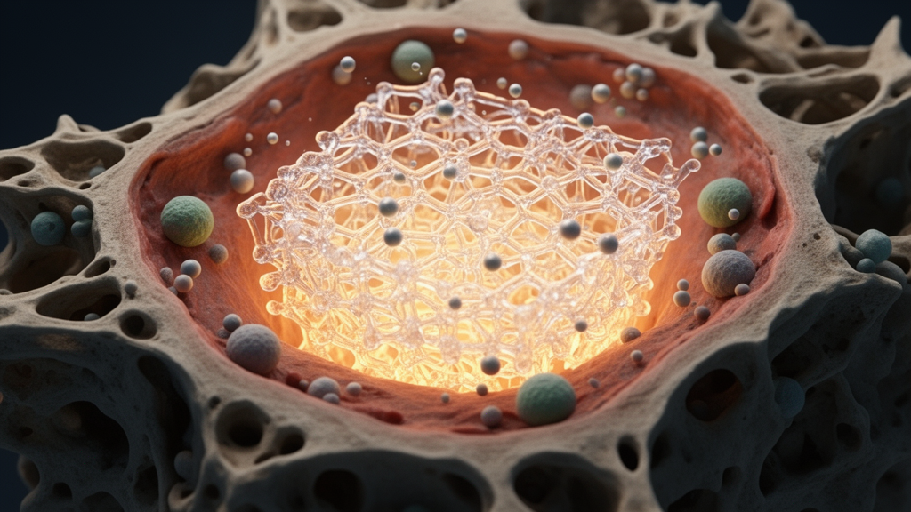
A comprehensive review published in Frontiers in Biomaterials Science by researchers from the Indian Institute of Technology Mandi examines the rapidly expanding landscape of smart hydrogel platforms engineered to address postoperative osteosarcoma. Osteosarcoma remains the most prevalent primary malignant bone tumor, characterized by aggressive local invasion and a notorious propensity for pulmonary metastasis, which serves as the principal cause of disease-related mortality. While current clinical protocols rely on a multimodal regimen of neoadjuvant chemotherapy, surgical resection, and postoperative adjuvant chemotherapy to target micrometastatic disease, patient outcomes drop drastically from a five-year survival rate of sixty to seventy-five percent in localized presentations to merely twenty to thirty percent once metastatic dissemination occurs. Furthermore, surgical intervention leaves behind critical-sized structural bone defects that cannot heal spontaneously, alongside residual microscopic tumor cells embedded within the compromised resection margins. In response to these complex therapeutic bottlenecks, researchers have increasingly investigated hydrogel-based biomaterials capable of providing localized, stimuli-responsive therapeutic delivery while concurrently establishing an osteoconductive matrix to facilitate functional skeletal regeneration.
The review provides a systematic analysis of literature published between 2015 and June 2026 across major indexing databases, analyzing the mechanistic distinctions, therapeutic outputs, and translational maturity of diverse hydrogel formulations. Conventional systemic chemotherapy, typically based on high-dose regimens of doxorubicin, cisplatin, methotrexate, and ifosfamide, frequently suffers from severe off-target systemic toxicity, poor local bioavailability, and the rapid emergence of multidrug resistance. In contrast, hydrogels offer a three-dimensional, highly hydrated polymeric architecture that closely mimics the natural extracellular matrix, exhibiting tunable mechanical compliance and the ability to conform seamlessly to irregular post-resection bone cavities. Unlike bioinert metallic implants that risk poor osteointegration, chronic inflammation, and stress shielding, hydrogels can be tailored as active local drug depots. In the pathological osteosarcoma microenvironment—which features acidic pH, elevated reactive oxygen species, dysregulated enzymatic expression, and altered vascular networks—smart hydrogels undergo reversible or irreversible physical and chemical shifts to trigger on-demand cargo release upon encountering endogenous pathological signals or non-invasive external stimuli such as light, temperature, ultrasound, and magnetic fields.
Investigating direct drug delivery approaches, the authors trace the transition from early passive depots to advanced multi-stimuli synthetic drug-eluting matrices. Synthetic chemotherapeutic formulations, such as galactose-based hydrogels functionalized with imatinib or visible light-cured glycol chitosan networks co-loaded with doxorubicin and cisplatin, have confirmed that sustained local matrix retention enhances malignant cell cytotoxicity compared with free systemic drugs. Recent platforms have integrated multimodal therapeutic strategies into injectable systems to prevent recurrence while supporting adjacent healthy bone tissue. For instance, injectable sodium alginate hydrogels loaded with two-dimensional titanium carbide MXene nanosheets, doxorubicin, and icariin combine localized near-infrared photothermal ablation with controlled chemotherapy, mitigating chemotherapy-induced osteogenesis impairment. Similarly, thermosensitive hydrogel platforms incorporating zinc oxide microspheres and polydopamine utilize mild photothermal heating alongside sustained zinc ion release to stimulate osteogenic differentiation, while magnetically guided biodegradable hydrogel microrobots carrying anlotinib and the MET inhibitor SCR1481B1 achieve active navigation and synergistic cytotoxicity to overcome targeted drug resistance in both two-dimensional and three-dimensional tumor cultures.
To overcome the intrinsic systemic toxicity and biological tolerance issues associated with synthetic chemotherapeutics, researchers have increasingly explored natural phytochemicals incorporated into stimuli-responsive hydrogels. Compounds such as curcumin, resveratrol, gambogic acid, and shikonin exhibit broad multi-targeted antitumor activity, anti-inflammatory characteristics, and lower baseline cytotoxicity toward healthy tissues. Recent preclinical studies detailed in the review highlight how formulation design dictates specific biological outcomes. An acid-responsive hydrogel co-delivering gambogic acid and calcium ions triggers mitochondrial apoptosis via intracellular calcium overload while preserving surrounding bone architecture and limiting pulmonary metastatic burden. In parallel, a shikonin-loaded nanocomposite hydrogel suppresses osteosarcoma cell proliferation via CDK1 inhibition while downregulating PD-L1 expression to reinvigorate local antitumor immunity. Similarly, an injectable copper-coordinated Tremella fuciformis polysaccharide hydrogel delivers pH-triggered copper release, inducing cuproptosis, cell cycle arrest, and mitochondrial damage in K7M2 osteosarcoma cells, while concurrently driving M1 macrophage polarization, enhancing CD8-positive T-cell infiltration, and upregulating essential osteogenic markers including alkaline phosphatase, osteopontin, osteocalcin, and Runx2.
Beyond bulk injectable matrices, microgels and nanogels have emerged as specialized three-dimensional nanoscale networks that provide superior injectability, deep tissue penetration, and high surface area for molecular encapsulation. Their diminutive dimensions permit precise chemical functionalization, enabling networks to undergo charge reversal, swelling, or covalent cleavage in response to the mildly acidic tumor microenvironment. Several systems exploit acid-labile Schiff base linkages formed between gelatin amine groups and oxidized polysaccharide aldehydes, which remain stable under physiological neutral conditions but degrade rapidly within acidic tumor margins to release encapsulated chemotherapeutics such as doxorubicin. Collagen-derived and gelatin-based nanogels further leverage natural matrix susceptibility to local collagenase enzymes, supporting continuous enzymatic erosion and sustained drug release. Biomimetic advances have also introduced cancer cell membrane-camouflaged nanogels, which demonstrate significantly enhanced homing and accumulation within target lesions compared with non-camouflaged carriers, alongside self-degradable hyaluronic acid nanogels that sequentially release hyaluronidase to degrade extracellular barriers before discharging intracellular cytotoxic proteins to induce targeted tumor cell destruction.
Despite the precise delivery capabilities of nanoscale carriers, their inherently soft structure and rapid clearance restrict their ability to withstand physiological forces in critical-sized, load-bearing skeletal defects. To bridge this divide, researchers have developed hydrogel-loaded composite platforms that incorporate reinforcing inorganic fillers, bioactive ceramics, and therapeutic nanoparticles. Formulations combining alginate, gelatin methacryloyl, or chitosan-polyacrylamide networks with bioglass, beta-tricalcium phosphate, or synthetic hydroxyapatite nanoparticles achieve superior structural stability while suppressing residual malignant recurrence. For example, magnesium nanoparticle-reinforced chitosan-polyacrylamide nanocomposite hydrogels provide durable photothermal ablation under near-infrared irradiation, completely preventing local tumor recurrence in animal models while sustained magnesium ion release drives mineralization, angiogenesis, and bone defect regeneration. Similarly, advanced composite constructs incorporate magnetic mesoporous bioactive glass or mineralized magnetic fibers, providing deep-penetrating magnetothermal or photothermal tumor eradication alongside long-term stem cell protection and calcium phosphate deposition, effectively synchronizing immediate oncological ablation with progressive structural skeletal repair.
Sustained therapeutic ion delivery represents another pivotal pillar of contemporary hydrogel engineering, harnessing bioactive inorganic ions to achieve simultaneous cytotoxicity and tissue reconstruction without relying solely on organic pharmaceuticals. Smart hydrogel depots engineered to release controlled amounts of zinc, strontium, magnesium, calcium, copper, manganese, gallium, selenium, and iron ions exert dual bioactivity. In these platforms, sustained ion discharge induces tumor eradication through mechanisms such as Fenton-like catalytic reactive oxygen species generation, cuproptosis, or interference with tumor iron metabolism, while modulating the immune microenvironment. Concurrently, ions like magnesium, zinc, and strontium upregulate osteogenic differentiation pathways and accelerate mineral deposition. For instance, organic-inorganic hydrogels integrating selenium- and magnesium-doped hydroxyapatite nanorods with calcium peroxide nanospheres have achieved complete tumor eradication alongside full structural bone defect healing over twelve weeks in vivo, demonstrating how ion release kinetics can be tailored to match the physiological timeline of bone repair.
The review also details how surface-functionalized and cell-laden hydrogel matrices advance biological mimicry beyond passive material cues. Functionalized hydrogels featuring catechol groups, oxidative stress-responsive linkages, or dynamic supramolecular networks have integrated immune checkpoint inhibitors, STING agonists, and cold atmospheric plasma-derived reactive species to stimulate immunogenic cell death and recruit dendritic cells. Meanwhile, cell-laden hydrogels incorporate patient-derived or representative cancer, stromal, and mesenchymal stem cells into tunable three-dimensional scaffolds. These models reproduce hypoxic gradients, nutrient transport limitations, and tumor-stroma crosstalk far more accurately than conventional monolayer cultures, demonstrating that stromal interactions directly elevate chemotherapeutic tolerance and cellular invasiveness. Embedded bioprinting strategies that generate biomechanical stiffness gradients within fibrin and decellularized extracellular matrix bioinks have replicated native tumor nests, migration dynamics, and drug resistance patterns, establishing advanced platforms for personalized therapeutic screening.
Advanced biofabrication technologies, specifically three-dimensional and four-dimensional bioprinting, represent an important frontier for fabricating patient-tailored osteosarcoma interventions. By integrating medical imaging data from computed tomography and magnetic resonance imaging, additive manufacturing enables the exact fabrication of patient-specific constructs that match irregular surgical resection margins. The emergence of four-dimensional bioprinting further incorporates temporal responsiveness, using stimuli-responsive bioinks such as silk methacrylate or modified alginate that undergo programmed shape-morphing or structural transformation over time upon encountering environmental cues like temperature or pH. Although 4D bioprinting has predominantly been evaluated in vascular and cartilage repair, the authors emphasize its immense potential for fabricating self-fitting, expanding implants capable of filling complex, non-uniform post-resection bone cavities while executing staged therapeutic delivery.
A critical contribution of the review is its rigorous evaluation of preclinical animal models used to assess hydrogel performance, identifying a substantial disconnect between laboratory testing and clinical disease reality. The majority of hydrogel studies continue to utilize small-animal murine models, predominantly relying on subcutaneous ectopic xenografts due to their simplicity and rapid establishment. However, subcutaneous sites completely fail to emulate the complex mechanical loading, cortical osteolysis, and immunological environment of native bone. Orthotopic murine models, established via intratibial or intrafemoral injection, provide improved bone-tumor interactions but remain constrained by small anatomical scale, altered immune profiles, and difficulties in surgically simulating wide tumor resection followed by local recurrence. Consequently, favorable outcomes achieved in rodent studies often fail to predict efficacy in human patients.
To establish a more realistic translational bridge, the authors highlight the necessity of intermediate and large animal models within the preclinical testing pipeline. Intermediate models, particularly orthotopic VX2 rabbit models involving long bones such as the femur or radius, successfully recreate characteristic pathological hallmarks including severe osteolysis, neoplastic calcification, load-bearing stress, and pulmonary micrometastases. When intermediate models are subjected to surgical resection and critical-sized defect creation, they consistently expose the primary mechanical failure of standalone hydrogels under true physiological loads, prompting the shift toward rigid ceramic-reinforced composite scaffolds. Furthermore, spontaneous canine osteosarcoma represents the most clinically representative large-animal model available, exhibiting natural genetic heterogeneity, native immune interactions, and clinical compatibility with limb-sparing surgical resection. Nevertheless, cost, experimental variability, and ethical hurdles have severely limited the evaluation of hydrogel systems in canine trials, leaving an unresolved validation gap prior to clinical translation.
In their final synthesis, the authors underscore that several decisive bottlenecks must be overcome before smart hydrogel systems can achieve clinical adoption. Most contemporary hydrogel research remains disproportionately focused on primary tumor ablation and localized bone repair, leaving pulmonary metastasis—the primary driver of patient mortality—largely unaddressed and unmonitored in experimental protocols. In addition, matching hydrogel degradation kinetics to the sequential phases of initial tumor eradication and prolonged osteogenesis remains technically challenging, while the introduction of complex multicomponent nanoformulations raises severe regulatory, reproducibility, and sterilization concerns. Overcoming these barriers will require interdisciplinary engineering strategies that combine image-guided fabrication, artificial intelligence, and machine learning to systematically optimize polymer crosslinking, degradation profiles, and multi-drug release kinetics. Ultimately, translating smart hydrogels from academic proof-of-concept constructs into clinically viable therapies demands standardized, mechanically rigorous post-resection models that validate sustained oncological control, functional load-bearing defect repair, and metastasis suppression simultaneously.
Subject of Research: Smart hydrogel biomaterials for localized postoperative osteosarcoma therapy and bone regeneration
Article Title: Smart hydrogel-based anticancer strategies for postoperative osteosarcoma management
Article References: Diwan, H., Behera, S. S., & Murab, S. (2026). Smart hydrogel-based anticancer strategies for postoperative osteosarcoma management. Frontiers in Biomaterials Science, 5, Article 1906677. https://doi.org/10.3389/fbiom.2026.1906677
Image Credits: AI Generated
DOI: 10.3389/fbiom.2026.1906677
Keywords: osteosarcoma, smart hydrogels, postoperative cancer management, bone defect repair, localized drug delivery, bioactive ions, composite hydrogels, nanogels, 3D bioprinting, photothermal therapy, chemodynamic therapy, doxorubicin, regenerative medicine, orthotopic animal models, preclinical evaluation, biomaterial scaffolds, cuproptosis, translational oncology
News Source: Nathaniel Bowman. (November 16, 2026). Review Evaluates Smart Hydrogels for Postoperative Osteosarcoma Recurrence and Regeneration. Scienmag.