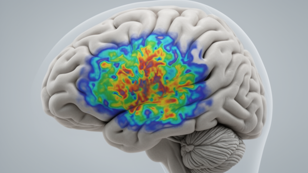
A prospective study of 94 preoperative patients with adult-type diffuse glioma has demonstrated that cellular microstructural quantification using time-dependent diffusion magnetic resonance imaging can noninvasively predict tumor grade and isocitrate dehydrogenase mutation status. Adult-type diffuse glioma represents the most common type of primary central nervous system tumor occurring in adults. Preoperative grading and molecular subtyping remain major clinical challenges, because surgical biopsy is invasive and cannot fully capture tumor heterogeneity across the entire lesion.
To address these clinical limitations and reduce dependency on invasive surgical procedures, researchers evaluated an advanced imaging approach that bridges the gap between macro-scale imaging and microscopic pathology. The investigative team implemented a 6.4-minute time-dependent diffusion magnetic resonance imaging protocol combined with biophysical modeling. Conducted on a 3.0-Tesla scanner with both pulsed and oscillating gradient diffusion sequences, this protocol functions as a clinically feasible virtual biopsy tool to evaluate tumor cell properties in patients prior to surgery.
Through biophysical modeling of the diffusion measurements, the researchers estimated several specific microstructural parameters characterizing tumor cell biology. These parameters included the intracellular volume fraction, designated as fin, the transcytolemmal water exchange time, denoted as τex, and apparent cellularity. In addition to these cellular parameters, the study evaluated conventional diffusion metrics, including standard apparent diffusion coefficient values, specifically ADC50ms, as well as the relative change in apparent diffusion coefficient across diffusion times, referred to as rcADC and cADC.
In diagnostic assessments for World Health Organization tumor grading, the estimated microstructural parameters demonstrated superior performance compared with conventional diffusion measurements. The intracellular fraction achieved the highest diagnostic accuracy, reaching an area under the receiver operating characteristic curve of 0.885. This was followed by the transcytolemmal water exchange time with an area under the curve of 0.864 and apparent cellularity with an area under the curve of 0.863. DeLong statistical testing showed that fin, τex, apparent cellularity, and rcADC all had significantly higher areas under the curve than the best conventional apparent diffusion coefficient, ADC50ms, which achieved an area under the curve of 0.739 with P values ranging from 0.002 to 0.027.
While fin, τex, and apparent cellularity performed well, other modeled parameters, specifically the cell diameter parameter d and the intracellular exchange lifetime τi, did not achieve significant diagnostic improvements over conventional diffusion measurements in grading. For the identification of isocitrate dehydrogenase mutation status, the intracellular fraction again yielded the highest diagnostic performance, attaining an area under the curve of 0.869. The transcytolemmal water exchange time and apparent cellularity also demonstrated strong diagnostic performance, yielding areas under the curve of 0.855 and 0.853, respectively. Both the intracellular fraction and apparent cellularity significantly outperformed ADC50ms in predicting mutation status.
To verify whether the noninvasive biophysical estimates accurately reflected microscopic cellular features, the authors performed histopathological validation using patient tissue samples. Microscopic evaluation of hematoxylin and eosin-stained tumor sections confirmed the physical relevance of the calculated imaging metrics. Specifically, histopathological analysis confirmed a statistically significant positive association between the magnetic resonance-derived intracellular fraction and the measured nuclear volume fraction on stained sections, yielding a correlation coefficient of r = 0.564 with a significance level of P < 0.0001.
The authors concluded that time-dependent diffusion magnetic resonance imaging microstructural quantification provides promising diagnostic value for noninvasive World Health Organization grading and isocitrate dehydrogenase mutation subtyping in adult-type diffuse glioma. Intracellular fraction and transcytolemmal water exchange time emerged as particularly valuable imaging biomarkers that outperform conventional apparent diffusion coefficient metrics and correlate with underlying histopathological features. By establishing accurate, pathology-correlated imaging biomarkers within a 6.4-minute acquisition protocol, this virtual biopsy approach demonstrates substantial clinical potential for precise preoperative diagnosis and neuro-oncological assessment.
Subject of Research: Noninvasive diffusion MRI microstructural quantification for diffuse glioma grading and molecular subtyping
Article Title: Cellular microstructural quantification predicts grading and IDH subtype in adult-type diffuse gliomas
Article References: Wang, S., He, J., Yang, S., Mao, J., Zhang, H., Lu, J., Li, H., Wei, K., Xu, H., Sun, J., Wu, D., & Wang, C. (2026). Cellular microstructural quantification predicts grading and IDH subtype in adult-type diffuse gliomas. BMC Medical Imaging. https://doi.org/10.1186/s12880-026-03058-9
Image Credits: AI Generated
DOI: 10.1186/s12880-026-03058-9
Keywords: adult-type diffuse glioma, time-dependent diffusion MRI, intracellular fraction, transcytolemmal water exchange time, apparent cellularity, IDH mutation status, WHO tumor grading, apparent diffusion coefficient, pulsed gradient diffusion, oscillating gradient diffusion, biophysical modeling, virtual biopsy, nuclear volume fraction, histopathology, 3.0T MRI scanner, preoperative neuroimaging, biomarkers, tumor heterogeneity
News Source: Ophelia Keating. (November 16, 2026). Diffusion MRI Microstructural Quantification Accurately Predicts Diffuse Glioma Grade and Subtype. Scienmag.