


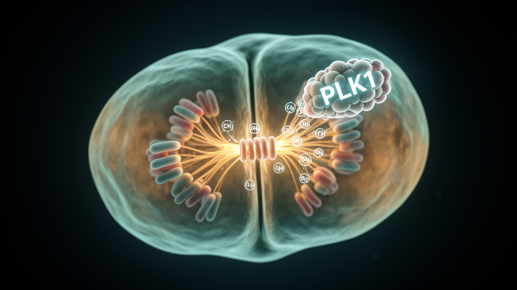
Few proteins sit closer to the heart of cell division than polo-like kinase 1, or PLK1. This highly conserved serine/threonine kinase reaches peak expression during the G2/M phase of the cell cycle and, through the phosphorylation of a broad spectrum of substrates, orchestrates nearly every stage of mitosis: entry into mitosis, centrosome maturation, spindle assembly, chromosome segregation and cytokinesis. A review published in Frontiers in Cell and Developmental Biology by Wenjing Li, Huiping Lin and Donghua Wen now pulls together what is known about how PLK1 itself is controlled, arguing that the kinase is governed not by a single molecular switch but by an interlocking network of post-translational modifications, or PTMs, whose crosstalk is only beginning to be mapped.
The authors frame their synthesis around five modifications that have been documented on PLK1: phosphorylation, ubiquitination, SUMOylation, methylation and O-GlcNAcylation. Each of these covalent tags can alter the kinase’s activity, protein stability, subcellular localization, substrate recognition or interactions with partner proteins. The central message of the review is that these modifications do not operate in isolation. Increasingly, the evidence shows them cooperating, antagonizing one another or acting in defined sequences, forming a dynamic regulatory network that fine-tunes PLK1 throughout the cell cycle and, when it goes wrong, in cancer.
Phosphorylation is the best-characterized layer of this network. The pivotal event is phosphorylation of threonine 210, widely recognized as the key step required for PLK1 activation. During G2 phase, CDK1 enhances the activity of the kinase Aurora A by promoting its association with the cofactor Bora, enabling Aurora A to phosphorylate PLK1 at T210 and trigger activation. In late G2, T210 phosphorylation also suppresses Bora-induced PLK1 dimerization, allowing the kinase to dissociate into its catalytically active monomeric form and license mitotic entry. As mitosis proceeds and Bora is degraded, checkpoint kinase 2 (CHK2) takes over, directly phosphorylating T210 to sustain PLK1 activity. Additional inputs come from VRK2, which phosphorylates T210 and blocks the ubiquitin ligase CHFR from tagging PLK1 for degradation; SHCBP1, which enhances Aurora A-dependent phosphorylation by binding the kinase’s polo-box domain; and RACK1, which recruits Aurora A to centrosomes to promote T210 phosphorylation during S phase. A separate, Aurora A-independent route runs through the PIM2–CHK1 cascade, in which PIM2 phosphorylates CHK1 at S280, enabling CHK1 to phosphorylate PLK1 at T210.
Genetic and functional studies have assigned distinct duties to the two major regulatory serine/threonine sites, T210 and S137. T210 phosphorylation is primarily tied to the G2/M transition, centrosome maturation, spindle assembly and regulation of the spindle assembly checkpoint. Phospho-mimetic T210D promotes mitotic entry and can partially rescue the G2 arrest caused by Cyclin A2 depletion, but it also induces checkpoint-dependent mitotic delay, while the phosphorylation-deficient T210A mutant produces monopolar spindles and mitotic arrest. Sustained T210-dependent signaling maintains chromosome alignment, segregation and cytokinesis, and supports spindle checkpoint integrity, chromosome stability and DNA replication initiation through the PLK1–MPS1 and PLK1–HBO1 pathways. S137 phosphorylation, detected in late mitosis, is associated with centrosome function and fine-tuning of mitotic progression: expression of the S137D mutant triggers untimely APC/C activation and frequent mitotic catastrophe, and in the DNA damage response CHK1-mediated S137 phosphorylation prevents PLK1 from being recruited to DNA double-strand breaks while preserving its kinase activity toward the repair protein RAD51.
Beyond these canonical sites, tyrosine phosphorylation adds a further layer of fine control. Four tyrosine sites have been identified: Y166, Y217, Y425 and Y445. PKMYT1 phosphorylates Y217, which suppresses T210 phosphorylation and dampens kinase activity. In contrast, the proto-oncogene kinase c-Abl phosphorylates Y425, Y217 and Y445, with Y425 predominant; Y425 phosphorylation induces a conformational change that strengthens the Aurora A–PLK1 interaction, promotes T210 phosphorylation and suppresses ubiquitin-dependent degradation, collectively accelerating mitotic entry. Reduced Y425 phosphorylation delays cell-cycle progression, and aberrant c-Abl-mediated phosphorylation has been associated with cervical cancer progression and poor prognosis. By contrast, c-Src-mediated phosphorylation of Y166 and Y445 suppresses PLK1 activity and is linked to impaired spindle checkpoint function and chromosome misalignment. Additional serine sites round out the picture: S99 phosphorylation, dependent on PI3K-AKT signaling, recruits 14-3-3γ and supports the metaphase-to-anaphase transition; MK2 phosphorylates S326, where disruption causes mitotic arrest; ERK phosphorylates T198, S404, S341 and S638, with S404 implicated in nuclear envelope integrity during embryonic development; and phosphorylation at T214, whose upstream kinase remains unknown, is required for normal mitotic function.
Counterbalancing all this activation, several phosphatases remove phosphate groups from PLK1. The MYPT1–PP1cβ complex is one of the best characterized: CDK1-dependent phosphorylation of MYPT1 promotes its binding to the PLK1 polo-box domain, recruiting PP1cβ to dephosphorylate T210 and suppress kinase activity. Upstream regulators including CHK1, CHK2, LATS1 and NUAK1 modulate this process by controlling MYPT1 phosphorylation, and O-GlcNAcylation of MYPT1 itself weakens its interaction with PLK1, thereby raising T210 phosphorylation. Protein phosphatase 2A, with its B55α and B55δ regulatory subunits, dephosphorylates and inactivates PLK1 after DNA damage, and during recovery from severe damage PP2A–B55-mediated dephosphorylation induces mitotic slippage, allowing compromised cells to exit mitosis without completing chromosome segregation. At kinetochores, PP1 and PP2A–B56 remove PLK1 from the BUB complex, terminating a positive-feedback loop and enabling spindle checkpoint silencing. PPP4C dephosphorylates S137 to support homologous recombination repair, PTEN restrains excessive phosphorylation of both Aurora A and PLK1, and the EYA1 and EYA4 phosphatases dephosphorylate Y445, restoring polo-box-mediated substrate binding and enabling centrosome maturation during G2.
Ubiquitination, the covalent attachment of ubiquitin by E3 ligases, controls both where PLK1 sits and how long it survives. The major ubiquitination sites identified so far are K492 and K209. CUL3–KLHL22-mediated mono-ubiquitination of K492 disrupts PLK1’s interaction with kinetochore phospho-receptors, releasing the kinase from kinetochores before sister chromatid separation without changing its overall abundance, a spatially restricted event essential for accurate chromosome alignment. Protein homeostasis is governed by several E3 ligases: SCFβTrCP targets PLK1 for degradation at the G1/S transition, APC/C-Cdh1 degrades it in G2 after DNA damage to prevent premature mitotic entry, UBAP2L removes PLK1 from mitotic structures, and the CUL4–CRBN complex degrades PLK1 in cells treated with immunomodulatory drugs in a p97-dependent manner. CHFR-mediated ubiquitination at K209 leads to PLK1 degradation, delayed CDC25C activation, sustained WEE1 activity and delayed mitotic entry, a pathway directly antagonized by VRK2-dependent T210 phosphorylation. Progressive proteasome-dependent degradation beginning at anaphase, requiring a D-box-like motif, permits CEP55 recruitment to the midbody and successful abscission. In tumors, ubiquitination can cut both ways: TRIM47-mediated K63-linked ubiquitination stabilizes PLK1 and activates NF-κB and MAPK signaling in hepatocellular carcinoma, while CHIP-mediated ubiquitination in prostate cancer participates in the DNA damage response, and its loss allows cells with unrepaired damage to continue cycling into mitotic catastrophe.
A family of deubiquitinating enzymes counteracts degradation, and the review catalogs a growing list of them: USP1, USP3, USP7, USP10, USP16, USP18, USP24, USP39 and OTUD3. Most stabilize PLK1 by removing K48-linked polyubiquitin chains, and many have been linked to malignant progression in specific cancers, including T-cell acute lymphoblastic leukemia, glioma, prostate cancer, pancreatic ductal adenocarcinoma, esophageal cancer, head and neck squamous cell carcinoma and gastric cancer, through effects on proliferation, glycolysis, oncogenic signaling such as mTORC1 and NOTCH1, and therapeutic resistance. USP16 plays a distinct spatial role, removing ubiquitin to promote PLK1 retention at kinetochores during early mitosis, forming a reversible cycle with CUL3–KLHL22 that controls kinetochore dynamics from prometaphase to metaphase. OTUD3, meanwhile, stabilizes PLK1 with neuroprotective effects in cerebral ischemia–reperfusion injury, acting through PI3K/AKT signaling and suppression of ferroptosis. Notably, deubiquitination itself is subject to PTM control: O-GlcNAcylation of USP16 enhances its activity toward PLK1, promoting faithful chromosome segregation.
The remaining modifications complete the network. Lysine methylation of PLK1, catalyzed by SETD6 at K209 and K413, SETD7 at K191 and EHMT2 (G9a) at K209, generally restrains kinase activity, slowing mitosis, limiting ATP consumption at kinetochores and coordinating DNA replication and repair. Strikingly, G9a-mediated methylation of K209 and Aurora A-mediated phosphorylation of T210 are mutually antagonistic, each modification preventing the other, creating a molecular switch that balances PLK1 activation. O-GlcNAcylation at T291, catalyzed by OGT, unusually promotes rather than protects against ubiquitination, driving PLK1 turnover; disruption of this site causes mitotic delay and chromosome segregation defects, and both T291A and T291N mutants promote uterine tumor growth in mouse xenograft models. SUMOylation at K492 promotes nuclear import and shields PLK1 from APC/C-Cdh1-mediated degradation, while in mouse oocytes PLK1 is modified by both SUMO-1 and SUMO-2/3 at distinct locations, and phosphorylated PLK1 at T210 and S137 colocalizes with the respective SUMO paralogs during meiosis II, suggesting functional cooperation in chromosome attachment and segregation. Dysregulated SUMOylation has also been associated with age-related meiotic defects in oocytes.
The authors are careful about what remains unknown. Only a limited number of PTM crosstalk mechanisms have been experimentally characterized, and key questions persist about the upstream enzymes involved, the temporal and spatial coordination of modifications, and how dysregulated PTM networks contribute to tumor progression and therapeutic resistance. They point to quantitative proteomics, structural biology, single-cell and spatial multi-omics and artificial intelligence-assisted protein modeling as tools expected to accelerate systematic mapping of the PLK1 PTM network. Because aberrant PLK1 expression is reported in prostate, ovarian, small-cell lung and pancreatic cancers and is frequently associated with aggressive disease and poor outcomes, a more complete picture of how its chemical tags interact could inform biomarker discovery and refine PLK1-targeted therapeutic strategies, though the review itself is a synthesis of existing literature rather than a demonstration of new clinical efficacy.
Subject of Research: Post-translational modifications of the mitotic kinase PLK1 and their crosstalk in cell-cycle regulation and tumor progression
Article Title: Post-translational modifications of PLK1: a dynamic regulatory network in cell cycle regulation and tumor progression
Article References: Li, W., Lin, H., & Wen, D. (2026). Post-translational modifications of PLK1: a dynamic regulatory network in cell cycle regulation and tumor progression. Frontiers in Cell and Developmental Biology, 14, Article 1987587. https://doi.org/10.3389/fcell.2026.1987587
Image Credits: AI Generated
DOI: 10.3389/fcell.2026.1987587
Keywords: PLK1, post-translational modifications, phosphorylation, ubiquitination, SUMOylation, methylation, O-GlcNAcylation, cell cycle, mitosis, tumor progression, cancer therapy, review
News Source: Nathaniel Bowman. (October 29, 2026). PLK1’s Hidden Code: How Chemical Tags Orchestrate Cell Division and Cancer. Scienmag.



