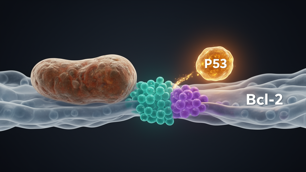
Scientists have mapped a new molecular battleground inside cutaneous squamous cell carcinoma (cSCC) cells, one of the most common forms of skin cancer. In a study published in the journal Cell Division, researchers report that the anti-apoptotic protein Bcl-2 promotes tumor cell survival by altering the function of mitochondria-associated membranes (MAMs), specialized contact sites between the endoplasmic reticulum and mitochondria. When the tumor suppressor p53 was overexpressed alongside Bcl-2, it reversed these effects, restoring calcium flux, mitochondrial membrane potential changes, reactive oxygen species production, and apoptotic signaling both in cultured cells and in mouse xenograft tumors.
The research team, led by Yupeng Huang, Jing Liu, Xiao Shao, Chunyan Xu, and Xianpeng Ma, constructed stable A431 cSCC cell lines using lentiviral transduction: one line overexpressing Bcl-2 alone, and another co-overexpressing Bcl-2 and p53. They then performed transcriptome sequencing (RNA-seq) to identify the downstream signaling changes, followed by pathway enrichment and protein-protein interaction network analyses. Functional assays including flow cytometry, CCK-8 viability testing, EdU incorporation, colony formation, and Transwell-based invasion assays quantified apoptosis, growth, migration, and invasive potential. Key MAMs functions—calcium flux, mitochondrial membrane potential, reactive oxygen species, and ATP levels—were measured directly, and subcutaneous tumor models in nude mice provided in vivo validation.
The RNA-seq analysis identified 560 differentially expressed genes in Bcl-2-overexpressing A431 cells, with 263 upregulated and 297 downregulated at a threshold of absolute log2 fold change greater than 1 and p less than 0.05. Gene Ontology enrichment showed that Bcl-2 overexpression predominantly affected mitochondrial outer membrane calcium transport and apoptotic signaling pathways, while Kyoto Encyclopedia of Genes and Genomes analysis pointed to the calcium signaling pathway and apoptosis-associated pathways. Protein-protein interaction network analysis, built on the STRING database with high-confidence interactions, showed that Bcl-2 directly regulated key apoptotic proteins including p53 (TP53), BAX, and PUMA (BBC3), and influenced MAMs-related proteins including VDAC2, IP3R (ITPR1), and SERCA2 (ATP2A2). Following Bcl-2 overexpression, the expression levels of all six of these genes were significantly downregulated.
Functionally, Bcl-2 overexpression markedly enhanced proliferation and migration of A431 cells while reducing apoptosis measured by flow cytometry and TUNEL staining. CCK-8 assays showed elevated viability, EdU staining and colony formation confirmed strengthened proliferative capacity, and wound-healing and Transwell assays revealed enhanced migration and invasion. Conversely, when the researchers knocked down Bcl-2 using shRNA, proliferation and invasion were suppressed and apoptosis was induced, supporting an oncogenic role for the protein in this cellular context. Critically, co-overexpression of p53 reversed the oncogenic phenotypes produced by Bcl-2: viability declined, proliferation weakened, migration and invasion decreased, and apoptosis was restored.
To test whether these findings generalized beyond a single cell line, the team repeated the key experiments in HSC-1, another human cutaneous squamous cell carcinoma cell line. Stable HSC-1 models overexpressing Bcl-2 alone or co-overexpressing Bcl-2 and p53 were generated, and target-gene expression was verified by RT-qPCR and Western blot. The functional results mirrored those in A431 cells: Bcl-2 overexpression suppressed apoptosis, increased proliferation, and promoted migration and invasion, while p53 co-expression effectively reversed these phenotypes. According to the authors, this consistency across two independent cSCC cell lines strengthens the reliability and generalizability of their conclusions.
The mechanistic core of the study lies in measurements of MAMs function. Using the Fluo-4 fluorescent indicator, the researchers found that free intracellular calcium was significantly reduced in Bcl-2-overexpressing cells compared with controls, whereas calcium levels rose significantly when p53 was co-overexpressed. JC-1 analysis demonstrated elevated mitochondrial membrane potential in Bcl-2-overexpressing cells, which fell significantly with p53 co-expression. The DCFH-DA fluorescent probe showed decreased intracellular reactive oxygen species with Bcl-2 overexpression and increased ROS after p53 co-overexpression, and ATP levels measured by the luciferin-luciferase method were higher with Bcl-2 overexpression and markedly reduced with p53 co-overexpression. Western blot analysis showed that the MAMs-associated proteins VDAC2, IP3R, and SERCA2, as well as the apoptotic markers Caspase-3, Bax, and PUMA, were downregulated by Bcl-2 overexpression and upregulated by p53 co-overexpression.
Co-immunoprecipitation assays verified a physical association between Bcl-2 and p53, with a significantly stronger interaction signal in Bcl-2-overexpressing cells and a markedly reduced signal in the co-overexpression group. Fluorescence colocalization experiments showed a significantly increased colocalization signal of the two proteins in Bcl-2-overexpressing cells, which was notably reduced when p53 was co-expressed. The same Co-IP and immunofluorescence co-localization results, along with consistent measurements of calcium, mitochondrial membrane potential, ROS, and ATP, were reproduced in HSC-1 cells. The authors interpret these data as evidence that Bcl-2 suppresses p53-mediated calcium transport and mitochondrial apoptosis signaling through MAMs, and that p53 can counteract this suppression.
In vivo validation used subcutaneous xenografts in BALB/c nude mice, with six mice per group inoculated with control, Bcl-2-overexpressing, or Bcl-2- and p53-co-overexpressing A431 cells. Serial caliper measurements showed that tumors in the Bcl-2 overexpression group grew significantly larger than controls, whereas tumors in the co-overexpression group showed a notable decrease in volume. On day 21, excised tumor masses were significantly increased in the Bcl-2 group and substantially reduced in the co-overexpression group. Hematoxylin and eosin staining showed increased cellular density and nuclear atypia in Bcl-2-overexpressing tumors, while co-overexpression tumors displayed expanded necrotic areas, disrupted tissue architecture, and prominent nuclear pyknosis. Immunohistochemistry confirmed higher Bcl-2 expression in the overexpression group and enhanced p53 staining in the co-overexpression group. In tumor tissues, ROS and ATP measurements and Western blots for VDAC2, IP3R, SERCA2, Caspase-3, Bax, and PUMA followed the same patterns observed in cultured cells.
The study’s context within cSCC biology is notable. Bcl-2 expression in clinical cSCC specimens is heterogeneous, with some cases showing high expression and others low or negative expression, and the authors emphasize that their gain-of-function strategy was designed to dissect mechanisms in the subset of cSCC characterized by functionally active Bcl-2 rather than to establish Bcl-2 as a universal driver. On the p53 side, genomic studies have shown that TP53 is the most frequently mutated driver gene in cSCC, with mutation rates of approximately 50 to 60 percent in primary tumors and as high as 85 to 95 percent in invasive or metastatic cases, most carrying ultraviolet-induced mutational signatures. MAMs themselves, membrane structures where the endoplasmic reticulum and mitochondria sit 10 to 30 nanometers apart, serve as central hubs for cellular calcium signaling, influencing mitochondrial membrane potential, ATP synthesis, and ROS production, and thereby modulating the balance between cell survival and apoptosis.
The authors acknowledge several limitations. The study relied primarily on in vitro cell experiments and subcutaneous xenograft models, lacking an in situ skin-derived cSCC model and validation in clinical tissue samples. The downstream signaling pathways were not comprehensively investigated, and the impact of different p53 mutational statuses on MAMs regulation was not explored. The authors suggest that future research should expand clinical sample sizes, employ techniques such as single-cell sequencing and live-cell imaging to analyze dynamic changes in MAMs, and explore combined therapeutic strategies targeting Bcl-2 inhibition, p53 activation, and MAMs modulation. They also note that potential resistance mechanisms, effects on normal tissues, and drug specificity would require thorough evaluation before any clinical translation of the proposed Bcl-2-MAMs-p53 signaling axis as a therapeutic target.
Subject of Research: How Bcl-2 and p53 regulate apoptosis through mitochondria-associated membranes in cutaneous squamous cell carcinoma
Article Title: p53 overexpression counteracts the pro-survival effect of Bcl-2 by restoring MAMs function in cutaneous squamous cell carcinoma
Article References: Huang, Y., Liu, J., Shao, X., Xu, C., & Ma, X. (2026). p53 overexpression counteracts the pro-survival effect of Bcl-2 by restoring MAMs function in cutaneous squamous cell carcinoma. Cell Division, 21(1), Article 18. https://doi.org/10.1186/s13008-026-00190-4
Image Credits: AI Generated
DOI: 10.1186/s13008-026-00190-4
Keywords: cutaneous squamous cell carcinoma, Bcl-2, p53, mitochondria-associated membranes, apoptosis, calcium signaling, RNA-seq, xenograft model, A431 cells, HSC-1 cells, tumor suppressor, mitochondrial membrane potential
News Source: Nathaniel Bowman. (October 29, 2026). p53 Reverses Bcl-2’s Pro-Survival Grip by Restoring MAM Function in Skin Cancer. Scienmag.