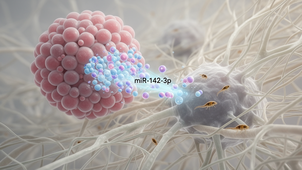
A 2026 review concludes that miR-142-3p has a dual role in cancer: it generally restrains malignant cells from within, yet its export in extracellular vesicles can give neighboring cells signals that favor tumor growth. Across lung, breast, cervical, colorectal, gastric, ovarian, glioma and leukemia models, the small non-coding RNA has been associated with lower proliferation, greater apoptosis, reduced invasion and improved sensitivity to chemotherapy. Once carried outside donor cells, however, the same molecule has been linked in limited experimental systems to cancer-associated fibroblast activation, angiogenesis and context-dependent changes in immune activity.
The evidence supports a model in which a molecule’s biological effect depends not only on its target genes, but also on where it is located, which cell receives it and how it is delivered. The review describes microRNAs, or miRNAs, as short, typically 18–22-nucleotide RNA molecules that regulate gene expression after transcription. They are transcribed as primary miRNAs and processed in the nucleus by the Drosha–DGCR8 Microprocessor complex, exported to the cytoplasm by exportin-5 and cleaved by Dicer into miRNA duplexes.
One strand is preferentially loaded into an Argonaute protein to form the RNA-induced silencing complex, which recognizes complementary response elements in target messenger RNAs and usually promotes their degradation or inhibits their translation. Canonical binding sites occur mainly in the 3-prime untranslated regions, although functional sites can also appear in the 5-prime untranslated region and coding sequences. Mature miRNAs and Argonaute-containing complexes can also enter the nucleus, where they may influence transcription-factor recruitment, DNA methylation, histone modifications and chromatin accessibility.
This regulatory system therefore has effects beyond the conventional cytoplasmic pathway. Within cancer cells, miR-142-3p is frequently reduced, and the available studies place this loss alongside more advanced disease or poorer treatment response. In lung adenocarcinoma, the review reports that expression was lower in approximately 52% of tumors than in adjacent normal tissue, while low levels were associated with larger tumors, advanced stage, lymph-node metastasis and treatment resistance.
Reduced expression has also been documented in breast, colorectal, gastric and ovarian cancer tissue or cell lines. Its effects are not universal, however, and the supporting experiments come from heterogeneous cancer models, including cultured cells and selected animal studies rather than a single large clinical cohort. The expression patterns also do not, on their own, establish the extent to which miR-142-3p loss causes tumor progression in patients.
One of the best-supported intracellular targets is high mobility group box 1, or HMGB1. In cervical cancer models, miR-142-3p bound the 3-prime untranslated region of HMGB1 messenger RNA, reduced HMGB1 expression and increased markers of apoptosis, including cleaved PARP, caspase-3, caspase-7, caspase-9 and BAX, while lowering anti-apoptotic Bcl-2 and Bcl-xL. Related suppressive effects were observed in glioma, where the RNA reduced cell viability and induced apoptosis through HMGB1 inhibition.
Other mechanisms include targeting CIAPIN1 in leukemia, Rac1/ERK signaling in colorectal cancer and FZD7 in cervical cancer, where FZD7 suppression inhibited oncogenic Wnt/beta-catenin signaling. These findings show that miR-142-3p can act through several context-dependent targets, but they do not establish that any single mechanism accounts for its behavior in human cancers. The molecule also appears to restrain epithelial–mesenchymal transition, a process in which cancer cells lose epithelial features and acquire properties associated with motility, invasion and dissemination.
In cervical cancer, increased miR-142-3p raised E-cadherin while reducing vimentin, N-cadherin, Snail and ZEB1, with corresponding reductions in migration and invasion. Suppression of FZD7 was linked to reduced beta-catenin nuclear translocation, while reduced NF-kappaB activity may also have contributed. Breast cancer experiments implicated CXCL12 and BACH1, and colorectal cancer experiments implicated Rac1/PERK signaling.
In lung cancer, miR-142-3p directly targeted NR2F6 and reduced MMP2 and MMP9; an MMP inhibitor abolished the migration induced by miR-142-3p knockdown. Most of these observations were generated in cell cultures, with some supporting animal experiments, so their clinical relevance remains incompletely defined. Chemosensitivity provides another major strand of evidence.
The review reports that miR-142-3p can enhance responses to agents including cisplatin, doxorubicin, docetaxel and paclitaxel in breast, lung and ovarian cancer models. In non-small-cell lung cancer, it targeted HMGB1, altered mTOR signaling and suppressed protective autophagy, increasing the effects of cisplatin and doxorubicin; tumors engineered to overexpress the RNA also responded more strongly to chemotherapy in vivo. In ovarian cancer, targeting SIRT1 sensitized resistant SKOV3 cells to cisplatin, and in H1299 lung cancer cells, SIRT1 suppression reduced autophagy and increased cisplatin sensitivity, with restoration of SIRT1 reversing the effect.
Other experiments linked miR-142-3p to apoptosis, stemness-associated proteins and reduced drug efflux. These are preclinical observations and do not establish that adding miR-142-3p would improve outcomes in treated patients. The review also notes that low intracellular miR-142-3p and higher levels in vesicles from chemoresistant cells have been observed relative to chemosensitive counterparts, but this does not prove that vesicle export causes resistance.
Extracellular vesicles are membrane-bound particles that transport proteins, lipids and nucleic acids between cells. Cancer cells can release more vesicles than nonmalignant cells, and therapeutic stress or drug resistance may increase release further. Limited evidence indicates that miR-142-3p is selectively enriched in tumor-derived vesicles: early lung adenocarcinoma cells produced vesicles containing more than four times the amount found in donor cells, and RNA-sequencing data from cervical cancer lines likewise showed higher extracellular than intracellular levels.
RNA-binding protein SYNCRIP recognizes a GGCU motif found in miR-142-3p and has been linked to sorting miRNAs into vesicles, offering a possible mechanism for this enrichment. Export could therefore lower a tumor cell’s internal tumor-suppressive activity while delivering a potentially supportive signal to recipients. The proposed link between export and intracellular depletion remains a mechanistic interpretation, because the review reports enrichment in selected models rather than a time-resolved demonstration in patients.
Recipient-cell evidence remains limited but is clearest for fibroblasts and blood vessels. In a lung adenocarcinoma model, vesicles transferred miR-142-3p to fibroblasts and increased canonical cancer-associated fibroblast markers, alpha-smooth muscle actin and PDGFR-beta, as well as migratory activity. Neither vesicle treatment nor direct overexpression significantly changed TGF-beta signaling, leaving the mechanism unresolved, and the result has not yet been demonstrated across tumor types or established in vivo.
For angiogenesis, miR-142-3p-enriched vesicles from oral cancer enhanced endothelial-cell angiogenesis in laboratory and animal experiments, while vesicles from regulatory T cells promoted proliferation, migration, cell-cycle progression and tube formation in human umbilical vein endothelial cells. Both settings implicated direct targeting of TGFBR1 and reduced TGF-beta/Smad signaling, potentially reducing anti-angiogenic mediators such as thrombospondin-1 and soluble Fms-like tyrosine kinase-1. A separate choroidal-neovascularization model showed that inhibiting miR-142-3p reduced vascular growth, but its relevance to cancer-associated angiogenesis was not tested.
Immune effects vary with cellular context and are not uniformly tumor-promoting. Overexpression in macrophages and dendritic cells was associated with reduced antigen processing, impaired antigen uptake and clearance, increased PD-L1 expression and lower production of the Th1-associated cytokines TNF-alpha and IFN-gamma; PD-L1 blockade restored T-cell proliferation in that experimental system. That study did not examine extracellular-vesicle delivery or cancer-specific immunity.
Conversely, vesicles from M1 macrophages carried miR-142-3p into glioblastoma cells, where the RNA suppressed HMGB1 and STAT3 signaling and reduced PD-L1, alleviating tumor-cell immune suppression. A separate hepatocellular carcinoma model linked intracellular miR-142-3p to suppression of ASH1L and increased immune activity, but did not test vesicle transfer. These studies used different sources, recipients and delivery mechanisms, leaving unresolved which variables determine whether the RNA strengthens or weakens antitumor immunity.
The most direct evidence for tumor-derived vesicles transferring the RNA into M1 macrophages came from hepatitis B virus-associated hepatocellular carcinoma, where the reported outcome was ferroptosis and tumor promotion; this is a specific experimental context rather than a general immune mechanism. The review also identifies cachexia as a hypothesis rather than a demonstrated cancer mechanism. Cancer-associated cachexia, characterized by weight loss and progressive skeletal muscle loss, affects an estimated 50–80% of patients with advanced disease and is associated with poorer survival and reduced treatment tolerance.
Direct evidence that tumor-derived vesicle miR-142-3p causes cachexia is absent. However, non-cancer experiments showed that miR-142-3p targets MEF2A in C2C12 myoblasts, disrupting cell-cycle progression and myotube formation; reducing miR-142-3p after nerve injury preserved muscle mass, strength, myofiber diameter and satellite-cell activity in mice. These findings make myogenic disruption a possible route to investigate, but they do not establish a role in cancer-associated cachexia.
Therapeutic development faces the central challenge that restoring miR-142-3p in tumor cells could restore growth suppression yet unintentionally support fibroblasts or endothelial cells if delivery is not precisely controlled. Mimics, chemically stabilized carriers, engineered vesicles and selective interruption of vesicle release or uptake are being investigated, but most programs remain preclinical, and available approaches may also affect physiologically important extracellular communication.
Subject of Research: Context-dependent tumor-suppressive and extracellular signaling functions of miR-142-3p
Article Title: miR-142-3p in cancer: intracellular tumor suppression and extracellular vesicle- mediated microenvironmental signaling
Article References: van der Merwe, M., Towle, R., Myburgh, K., Garnis, C., & Engelbrecht, A.-M. (2026). miR-142-3p in cancer: intracellular tumor suppression and extracellular vesicle- mediated microenvironmental signaling. Molecular Biology Reports, 53(1), Article 1501. https://doi.org/10.1007/s11033-026-12631-y
Image Credits: AI Generated
DOI: 10.1007/s11033-026-12631-y
Keywords: miR-142-3p, cancer biology, extracellular vesicles, tumor microenvironment, chemotherapy resistance, angiogenesis, cancer-associated fibroblasts, immune modulation, cachexia, microRNA therapeutics, cancer, intracellular
News Source: Nathaniel Bowman. (October 22, 2026). miR-142-3p suppresses tumors but can reshape their surroundings. Scienmag.