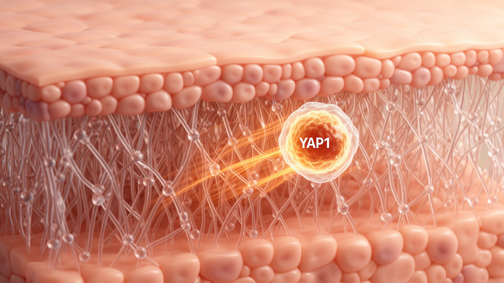
Keratin 9, a structural protein concentrated in the skin of palms and soles, also acts as a regulator of mechanical signalling in palmoplantar epidermis, according to a study combining human skin samples, mouse models, engineered keratinocytes and compression experiments. The researchers found that keratin 9 promotes the cytoplasmic retention of YAP1, a transcriptional effector of mechanical cues, while disease-associated mutations disrupt this function. Loss of keratin 9 produced abnormally nuclear YAP1, elevated keratin 16 and thickened palmoplantar epidermis; conversely, blocking YAP1 activity reduced the resulting lesions in mice. The work provides a mechanistic explanation for how mechanical stress may help establish the specialised identity of palmoplantar skin and links dysregulated YAP1 signalling to palmoplantar epidermal differentiation disorders, including KRT9-pEDD.
Human punch biopsies were used to compare healthy trunk and plantar skin with lesional plantar skin from patients carrying KRT9 variants associated with palmoplantar epidermal differentiation dysplasia, or KRT9-pEDD. Immunofluorescence revealed that healthy differentiating epidermal cells generally contained weak, diffuse cytoplasmic YAP1, with a small population of YAP1-positive nuclei. In KRT9-pEDD lesions, by contrast, strong nuclear YAP1 was common in the suprabasal epidermis. The population of keratin 14-negative, YAP1-positive nuclei increased by almost tenfold, and these nuclei extended further from the basal layer than in healthy skin. The affected epidermis also had significantly thicker suprabasal layers. At the individual-cell level, keratin 9 intensity was negatively correlated with nuclear YAP1 in healthy plantar skin but positively correlated in patient lesions, indicating that the relationship between the two proteins was reversed in disease.
Keratin 16, a stress-associated keratin normally present in palmoplantar epidermis, was also elevated in KRT9-pEDD samples. Healthy plantar cells showed a negative relationship between keratin 9 and keratin 16 abundance, whereas no meaningful relationship was detected in lesional cells, where keratin 16 remained high regardless of the amount of keratin 9 present. Single-cell RNA sequencing likewise showed opposing expression patterns in healthy skin. Transcription and protein data further indicated that the strongest keratin 9-expressing cell population was enriched for cornified-envelope formation, programmed cell death and keratinocyte differentiation, while the strongest keratin 16-expressing population was enriched for innate immune activation and wound-healing functions. These observations linked abnormal YAP1 partitioning with a shift away from the normal differentiation programme in KRT9-pEDD tissue.
The same molecular pattern emerged in Krt9-null mice. Wild-type and knockout newborn paws had similar YAP1 localisation at postnatal day 0, but by day 3 the knockout epidermis displayed abnormal nuclear YAP1 in suprabasal layers. The phenotype became more pronounced at 3.5 weeks, when palmoplantar lesions were already apparent. Total YAP1 abundance did not differ significantly between genotypes, but the ratio of phosphorylated, cytoplasm-retained YAP1 to total YAP1 was reduced in knockout footpads by day 3. The 14-3-3σ protein, which binds phosphorylated YAP1, was also elevated. RNA sequencing identified increased expression of the previously described YAP1 target Krt19 at days 0 and 3, while keratin 16 was elevated by day 3. The early onset of these changes indicates that abnormal YAP1 partitioning precedes visible keratoderma in this model.
Protein analysis during normal mouse development revealed a rapid transition among palmoplantar keratins. Quantitative mass spectrometry of whole paws from embryonic day 18.5 through postnatal day 10 showed that five intermediate filament proteins changed dynamically: keratins 16, 6B, 17, 2 and 9. Their expression shifted from keratin 6 and 16 dominance at days 0–1 to keratin 2 and 9 dominance at days 3–10. RNA sequencing showed that the corresponding transcript changes preceded the protein transition. This sequence coincides with the period when young paws begin encountering compression through nursing and early locomotion, although the experiments do not directly establish which specific postnatal behaviour generated the signal.
The researchers tested whether the keratin transition was dependent on YAP1 transcriptional activity using a mouse line in which a synthetic inhibitor of TEAD proteins could be induced in basal epidermal keratinocytes. Blocking YAP1 binding to TEAD reduced keratin 16 protein and messenger RNA at postnatal day 1 and delayed the subsequent rise in keratin 9 transcript at day 2. Chromatin immunoprecipitation in post-confluent N/TERT2G keratinocytes found YAP1 bound to two of three candidate TEAD sites within 1 kilobase upstream of the KRT16 transcription start site, but not to the tested site upstream of KRT10. These results support direct YAP1 regulation of keratin 16 and show that the early keratin 16 increase is needed, at least in part, for normal timing of keratin 9 expression.
Compression experiments provided direct evidence that mechanical force can induce palmoplantar-associated keratins. N/TERT keratinocytes were embedded in an agarose and type I collagen hydrogel designed to approximate the mechanical properties of viable plantar epidermis, then exposed to 10 kilopascals of static compression for 24 hours. Compression increased transcripts for keratin 9, keratin 17 and keratin 16, along with the canonical YAP1 target CYR61. These responses were absent when the cultures were treated with 2.5 micromolar verteporfin, which was used to inhibit YAP1 transcriptional activity. A431 epidermal carcinoma cells showed the same compression-dependent induction of keratins 9, 16 and 17 and CYR61. Knocking out KRT16 prevented compressed N/TERT cells from increasing KRT9, even though actin and keratin 6 responses remained, indicating that keratin 16 is required for the compression-induced expression of keratin 9 in this cultured system.
Cell-based experiments identified a mechanism through which keratin 9 restrains YAP1. Expression of wild-type keratin 9 in HeLa cells reduced the nuclear-to-cytoplasmic ratio of YAP1 and lowered transcription of CYR61. Two KRT9-pEDD-associated variants, R163Q and C406A, failed to produce either effect. Wild-type keratin 9 also physically interacted with 14-3-3σ in co-immunoprecipitation experiments, and proximity assays showed a similar degree of association between wild-type keratin 9 and 14-3-3σ as between keratin 14 and 14-3-3σ. The two pathogenic variants significantly reduced that association. By contrast, keratin 16 did not retain YAP1 in the cytoplasm despite sharing a conserved stutter cysteine with keratins 9 and 14. The findings support a model in which keratin 9, through 14-3-3σ, forms part of a complex that sequesters YAP1 in the cytoplasm and limits its transcriptional activity.
Topical verteporfin partially reversed the disease-associated phenotype in 3.5-week-old Krt9-null mice. One paw received 3 milligrams per millilitre verteporfin and the contralateral paw vehicle for seven days. Relative to vehicle, treatment reduced the thickness of the living epidermal layers by an average of 40 ± 18.9% and lowered keratin 16 transcript and protein. The fraction of epidermal nuclei containing YAP1 fell to levels observed in wild-type mice, YAP1-positive nuclei shifted closer to the basement membrane, and the phosphorylated-to-total YAP1 ratio increased. Genetic inhibition produced a similar result: inducing a TEAD inhibitor in basal keratinocytes of Krt9-null mice restored living-layer thickness to levels of control animals and significantly reduced keratin 16 staining. The study therefore identifies YAP1 activity as a contributory cause of the mouse lesions rather than merely a correlate, although verteporfin can affect processes beyond YAP1 and the genetic experiment provides the more specific intervention.
The results extend beyond KRT9-pEDD because variants in more than 50 genes can cause disorders of palmoplantar epidermal differentiation, and some affected individuals have reduced keratin 9 expression. The authors propose that several forms of palmoplantar keratoderma may converge on inadequate keratin 9, abnormal YAP1 activity and excessive keratin 16. The research nevertheless has important limits. The human analysis included a small number of patient samples, the principal mechanistic work used cultured cell lines and engineered keratinocytes, and the mouse model reproduces features of KRT9-pEDD rather than the complete human disorder. The experiments also do not yet explain how mechanically induced keratin 16 triggers keratin 9 or precisely how phosphorylation and 14-3-3 proteins establish the keratin-dependent YAP1 complex. Nevertheless, the combined evidence shows that keratin 9 is not only a mechanical scaffold but a signalling regulator, and that controlling YAP1 could potentially reduce palmoplantar thickening in a subset of inherited skin disorders. No clinical effectiveness or safety in patients was established by the work.
Subject of Research: Keratin 9 regulation of YAP1 signalling in palmoplantar epidermal differentiation and KRT9-pEDD
Article Title: Keratin 9 regulates mechanosensitive YAP1 signalling in palmoplantar epidermis
Article References: Steiner, S. N., Horst, E. N., Chiu, E. S., Athaiya, M., Johnson, C. N., Shen, J. Y., Kerns, M. L., Kirma, J. D., McCarthy, R. L., Shah, S. F. H., Sarig, O., Samuelov, L., Sprecher, E., O’Toole, E. A., Gudjonsson, J. E., Mehta, G., Iglesias-Bartolome, R., & Coulombe, P. A. (2026). Keratin 9 regulates mechanosensitive YAP1 signalling in palmoplantar epidermis. Nature Cell Biology. https://doi.org/10.1038/s41556-026-02087-4
Image Credits: AI Generated
DOI: 10.1038/s41556-026-02087-4
Keywords: keratin 9, YAP1 signalling, palmoplantar epidermis, KRT9-pEDD, mechanotransduction, keratin 16, skin differentiation, keratoderma, 14-3-3σ, mechanical compression, Keratin, regulates
News Source: Drew Townsend. (October 22, 2026). Keratin 9 controls mechanical YAP1 signalling in palmoplantar skin. Scienmag.