In a study published in Frontiers in Molecular Neuroscience, researchers report that lncR-AK158273 suppresses microglial autophagy in LPS-induced neuroinflammation models by upregulating Mst1 expression. The study, authored by Yongcang Zhang, Xinyan Fu, Lixia Guo, Xinfang Liu, Liang Tang and Lan Liu, with corresponding author Lan Liu at Xizang University and Liang Tang at Changsha Medical University, reports that this RNA molecule suppresses the cellular recycling process in microglia by boosting levels of Mst1, a kinase encoded by the STK4 gene.
The study used an LPS-NCI mouse model and an LPS-stimulated BV2 microglial cell model. In mice, they injected LPS directly into the lateral ventricles to create what the authors call an LPS-induced neuroinflammation cognitive impairment model, or LPS-NCI. Eighteen male Kunming mice, six months old and weighing roughly 26 grams, were divided into three groups of six: one receiving LPS, one receiving saline as a control, and a sham group that underwent surgery with holes drilled but no injection. Behavioral testing was performed on day 8 using the Y-maze test and on days 9-10 using the novel object recognition test. The Y-maze test, which measures spontaneous alternation as mice explore three arms, and the novel object recognition test, which tracks how long animals investigate a new object versus a familiar one, both revealed significant deficits in the LPS-treated animals compared with controls and shams.
Biochemical and histological analyses showed that the behavioral deficits were accompanied by elevated CD11b, TNF-α, IL-6, p-tau, and Aβ1-42, and by neuronal damage in the hippocampal CA1 region. Levels of CD11b, a marker of activated microglia, rose sharply in the hippocampus, as did the pro-inflammatory cytokines TNF-α and IL-6 measured by ELISA. Nissl staining of the hippocampal CA1 region, a subfield particularly vulnerable to inflammatory injury, showed a higher density of damaged neurons characterized by shrunken cell bodies, pyknotic nuclei, and dissolved Nissl bodies. The researchers also detected elevated levels of phosphorylated tau and amyloid-beta 1-42, two proteins closely associated with neurodegenerative pathology, while total tau remained unchanged across groups.
The critical molecular finding came when the team measured lncR-AK158273 and STK4 expression. Both were significantly upregulated in the hippocampi of LPS-NCI mice and in BV2 microglial cells exposed to LPS in culture.
To examine the role of lncR-AK158273 in autophagy, the researchers transfected si-lncR-AK158273 or pcDNA-lncR-AK158273 into LPS-stimulated BV2 cells. When they transfected cells with a plasmid to overexpress lncR-AK158273, they observed a cascade of changes consistent with autophagy suppression: Beclin-1 protein dropped, the ratio of LC3II to LC3I fell, and P62, a cargo receptor that accumulates when autophagic clearance stalls, rose. Conversely, silencing lncR-AK158273 with a specific siRNA produced the opposite pattern, restoring Beclin-1 and LC3II/LC3I while reducing P62. Transmission electron microscopy provided visual confirmation, showing fewer autophagosomes per cell in the overexpression group and more in the knockdown group.
Autophagic flux was determined using mRFP-GFP-LC3 fluorescence tracing with chloroquine treatment to block lysosomal degradation. After chloroquine treatment, yellow autophagosomes accumulated in the control + CQ group, which the authors state validated the inhibitory effect of chloroquine on lysosomal degradation. In control cells treated with chloroquine, this accumulation was dramatic. The authors interpret the lack of difference in autophagosome counts between the LPS group and the LPS plus chloroquine group as evidence that LPS suppresses autophagy primarily by preventing autophagosome biogenesis, not by impairing lysosomal degradation.
The causal chain linking lncR-AK158273 to Mst1 was then tested directly. Overexpressing the lncRNA in LPS-treated BV2 cells significantly increased both STK4 mRNA and Mst1 protein, while knocking down the lncRNA reduced both. Rescue experiments sealed the argument: when the researchers overexpressed Mst1 alone, autophagy markers shifted toward suppression, and silencing lncR-AK158273 partially reversed this effect. Conversely, when they knocked down Mst1, autophagy increased, and overexpressing lncR-AK158273 partially blunted that enhancement.
Immunofluorescence co-localization staining in mouse hippocampal tissue revealed that the autophagic defects were cell-specific. LC3 signals, a marker of autophagosomes, were predominantly localized within CD11b-positive microglia in the LPS-NCI group, with no obvious alterations observed in neurons or astrocytes.
The authors are candid about the study’s limitations. Only male mice were used, a choice made to avoid variability from estrous cycling but one that introduces sex bias, since microglial activation and inflammatory responses are known to differ between sexes. The LPS model represents acute exogenous inflammation rather than the chronic low-grade inflammation characteristic of human aging-related diseases. Nissl and hematoxylin-eosin staining cannot provide precise quantification of neuronal damage, and unbiased stereology would be needed for more accurate three-dimensional counts. The BV2 cell line, while a classic microglial model, cannot fully substitute for primary microglia. The precise molecular mechanism by which lncR-AK158273 regulates Mst1, whether through microRNA sponging, RNA-protein interaction, transcriptional activation, or chromatin modification, remains unresolved.
The authors state that the findings suggest a potential molecular target for central nervous system diseases related to neuroinflammation. Prior research cited by the authors reported that LPS binds to TLR4 and inhibits the FOXO3 transcription factor, downregulating autophagy-related gene expression, and that reduced LAMP2A leads to p300 accumulation, promoting NF-κB transcriptional activity and NLRP3 inflammasome activation.
Subject of Research: Mechanism by which lncRNA AK158273 regulates autophagy in LPS-induced neuroinflammation through Mst1 upregulation in microglia
Article Title: LncRNA‑AK158273 suppresses lipopolysaccharide‑triggered autophagy by up‑regulation of Mst1 expression in microglia
Article References: Zhang, Y., Fu, X., Guo, L., Liu, X., Tang, L., & Liu, L. (2026). LncRNA‑AK158273 suppresses lipopolysaccharide‑triggered autophagy by up‑regulation of Mst1 expression in microglia. Frontiers in Molecular Neuroscience, 19, Article 1973037. https://doi.org/10.3389/fnmol.2026.1973037
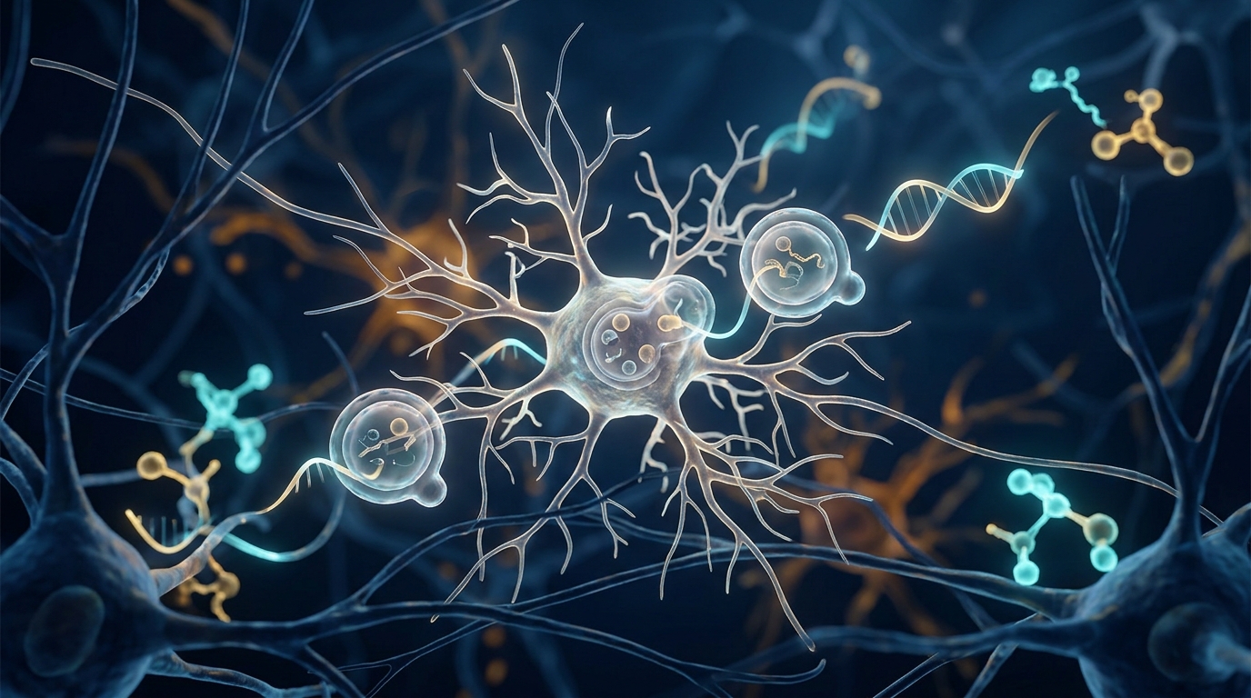
Image Credits: AI Generated
DOI: 10.3389/fnmol.2026.1973037
Keywords: lncRNA AK158273, Mst1, STK4, autophagy, microglia, neuroinflammation, LPS, BV2 cells, hippocampus, cognitive impairment, TLR4, Frontiers in Molecular Neuroscience
News Source: Cassandra Pierce. (November 4, 2026). LncRNA AK158273 curbs microglial autophagy via Mst1 in LPS neuroinflammation model. Scienmag.