A cadmium-based crystal grown from a simple water solution has shown stronger killing of two human cancer cell lines than the platinum drug cisplatin in laboratory tests, according to a study published in Results in Chemistry. Researchers grew single crystals of guanidinium cadmium chloride, a compound combining a guanidinium cation with a cadmium chloride anion, and then exposed A549 lung cancer cells and MCF-7 breast cancer cells to the material. In MTT assays, GuCC reduced the viability of both cell lines in a concentration-dependent manner, with lower IC50 values than cisplatin, the reference drug used in the comparison, and it also showed free-radical scavenging activity in DPPH, ABTS+, nitric oxide, and superoxide assays with mixed results against the ascorbic acid standard.
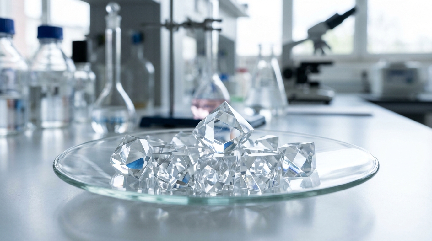
The compound, abbreviated GuCC by the authors, was prepared by dissolving guanidinium carbonate and cadmium chloride in distilled water in a molar ratio of 1:2. The two precursor solutions were mixed under continuous magnetic stirring for four hours, filtered to remove insoluble impurities, and left at room temperature for slow evaporation. After three weeks, single crystals suitable for analysis had formed.
Single-crystal X-ray diffraction revealed that GuCC crystallizes in the monoclinic crystal system with space group P21/c. The unit cell measures a = 3.8194(4) Å, b = 18.9724(13) Å, and c = 10.5067(8) Å, with a volume of 761.00(11) ų and four formula units per cell. The empirical formula is (CH6N3)+·(CdCl3)−, with a formula weight of 278.84. The structure was solved and refined using the SHELXL program, and a low R-factor of 2.55 percent for reflections with I greater than 2σ(I) confirmed the quality of the refinement. The crystallographic information has been deposited with the Cambridge Structural Database under CCDC number 2,286,602.
The asymmetric unit contains half of a metal-halide octahedron, formed by one cadmium atom and three chloride atoms, together with one guanidinium cation. Cadmium sits at the center of an octahedral site surrounded by six chlorine atoms, with metal-halide bond distances ranging from 2.518(1) to 2.762(1) Å. The guanidinium cation and the metal-halide polyhedral anion are connected through two three-centered and four two-centered classical N–H···Cl hydrogen bonds. These interactions generate four distinct ring motifs, labeled R1²(4), R2¹(6), R2²(8), and R4⁴(10), which link adjacent polyhedra and extend the crystalline framework. Octahedra share faces through bridging chloride atoms, forming chains parallel to the a-axis of the unit cell.
Thermogravimetric analysis carried out from 30 to 900 °C under nitrogen showed that the material remains thermally stable up to 311 °C. Decomposition then proceeds in two main stages: an initial weight loss of 34.3 percent between 311 and 386 °C attributed to liberation of amine groups, followed by a second loss between 386 and 693 °C as remaining organic moieties are removed. The final residue consists of carbon, nitrogen, cadmium, and chlorine.
To explore possible biological targets, the team performed molecular docking of GuCC against six proteins associated with breast and lung cancer: AKT1, BRCA1, and caspase-6 for breast cancer, and CDK2, EGFR, and KRAS for lung cancer. Predicted binding affinities ranged from −4.8 to −5.9 kcal/mol, with the strongest scores of −5.9 kcal/mol for caspase-6 and CDK2. Docking of the ligand into AKT1 involved hydrogen bonds with GLU A:298 and ASP A:274, while interactions with BRCA1 included contacts with ALA X:1708, LEU X:1786, TYR X:1845, CYS X:1759, and SER X:1755. For caspase-6, hydrogen bonds formed with ARG A:64 and SER A:120. In lung cancer targets, CDK2 bound through GLY C:16 and GLU C:12, EGFR through ASN A:818 and PHE A:832, and KRAS through GLN A:150. The docking protocol was validated by re-docking co-crystallized ligands, yielding RMSD values of 0.742 Å for AKT1, 1.768 Å for CDK2, 0.948 Å for EGFR, and 1.132 Å for KRAS, all below the 2.0 Å threshold commonly used to confirm docking reliability. Re-docking was not possible for caspase-6 and BRCA1 because their chosen crystal structures lack co-crystallized ligands. The authors emphasize that these docking results are computational predictions of binding, not proof of biological activity or mechanism.
The central biological finding came from MTT assays on A549 and MCF-7 cells. After 24 hours of exposure, GuCC reduced viability of both cell lines in a concentration-dependent manner. The IC50 value, the concentration that inhibits half of the cells, was 14.33 ± 0.93 μg/mL for A549 lung cancer cells and 12.16 ± 1.21 μg/mL for MCF-7 breast cancer cells. Cisplatin, tested in parallel as a reference, required higher concentrations to achieve the same effect, with IC50 values of 27.05 ± 0.46 μg/mL for A549 and 23.72 ± 1.64 μg/mL for MCF-7. All experiments were run in triplicate, and differences between treated and control samples were assessed with one-way ANOVA.
Morphological and fluorescence staining assays pointed toward apoptosis as the mode of cell death. When cells were treated with the IC50 concentration of GuCC for 24 hours, microscopic examination showed nuclear shrinkage and cell detachment, signs of cellular damage consistent with apoptotic death. Acridine orange–ethidium bromide double staining, which distinguishes live cells with intact membranes from apoptotic and dead cells, showed that treated A549 and MCF-7 cells fluoresced red or orange rather than the green of intact living nuclei, indicating loss of membrane integrity and chromatin condensation. DAPI staining, which binds adenine-thymine clusters in the minor groove of double-stranded DNA and highlights compacted nuclei, revealed condensed, horseshoe-shaped, and fragmented nuclei in treated cells.
Beyond the anticancer results, GuCC showed free-radical scavenging activity in DPPH, ABTS+, nitric oxide, and superoxide assays, with performance that varied against the ascorbic acid standard: weaker in the DPPH and ABTS+ assays but stronger in the nitric oxide assay. In the DPPH assay, GuCC had an IC50 of 42.18 ± 1.58 μg/mL, higher than the ascorbic acid control at 7.98 ± 0.96 μg/mL, indicating moderate activity by that measure. Nitric oxide scavenging was stronger: GuCC reached an IC50 of 35.65 ± 0.47 μg/mL, outperforming the standard at 52.08 ± 0.59 μg/mL. The ABTS+ assay yielded an IC50 of 62.15 ± 0.78 μg/mL, and superoxide radical scavenging gave 51.09 ± 0.65 μg/mL.
Important caveats remain: the cytotoxicity data come from two cancer cell lines in culture, the docking scores are computational predictions, and the antioxidant measurements are chemical assays rather than demonstrations of activity in organisms. Whether GuCC can selectively kill tumor cells in a living system, what its toxicity profile would be in healthy tissue, and how a cadmium-containing compound could be safely formulated are questions that will require animal studies and, ultimately, clinical evaluation before any therapeutic application can be considered.
Subject of Research: Synthesis, crystal structure, and in vitro anticancer and antioxidant evaluation of guanidinium cadmium chloride in A549 and MCF-7 cell lines
Article Title: Structural elucidation and biological evaluation of guanidinium cadmium chloride in A549 and MCF-7 cell lines with enhanced antioxidant activities
Article References: Siva, V., Murugan, A., Shameem, A. S., Kalaiarasi, G., Athimoolam, S., Viswanathan, T. M., Jauhar, R. M., & Peera, S. G. (2026). Structural elucidation and biological evaluation of guanidinium cadmium chloride in A549 and MCF-7 cell lines with enhanced antioxidant activities. Results in Chemistry, 32, Article 104015. https://doi.org/10.1016/j.rechem.2026.104015
Image Credits: AI Generated
DOI: 10.1016/j.rechem.2026.104015
Keywords: guanidinium cadmium chloride, A549 lung cancer cells, MCF-7 breast cancer cells, cisplatin, MTT assay, apoptosis, molecular docking, single-crystal X-ray diffraction, antioxidant activity, DPPH assay, hydrogen bonding, metallodrugs
News Source: Nathaniel Bowman. (November 5, 2026). Guanidinium Cadmium Chloride Crystal Outperforms Cisplatin in Lab Cancer Cell Tests. Scienmag.