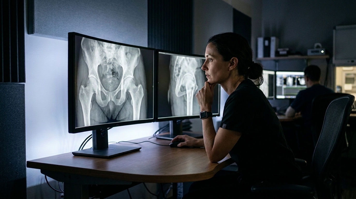
Researchers at the University of Oxford, working with a specialist from Alder Hey Children’s Hospital in Liverpool, have developed a method that lets an artificial intelligence model express how confident it is when it measures a baby’s hip from an ultrasound image. The work, published as an original research article in Frontiers in Medical Technology, targets a screening task performed on newborns: checking for developmental dysplasia of the hip. Its central claim is unusual for medical AI. Rather than only asking whether the model gets the measurement right, the team built a way for the model to signal when the measurement itself is uncertain.
The problem begins with how clinicians read these images. Ultrasound assessments of the infant hip depend on anatomical landmarks, specific points on the image that a clinician marks by hand. From those points, the clinician derives standard measurements, either an angle or a distance. In hip screening, two measurements dominate: the Graf alpha angle, used to assess how well the socket of the hip joint is formed, and the femoral head coverage percentage, which describes how much of the femoral head sits within the socket. Screening pathways then apply predefined thresholds to these numbers, classifying each infant as healthy or unhealthy.
That pipeline has a weak point, and it is human. Clinicians do not place landmarks identically. Natural variability exists among clinicians in where the landmark is placed, and because the measurement is computed from the landmark, the derived number and even the screening decision can shift with it. The authors describe this as genuine uncertainty in the assessment, not a mistake to be eliminated but a real property of the task. A measurement that sits near a threshold may flip from healthy to unhealthy depending on small differences in landmark placement, and different experienced readers can legitimately disagree.
The new method tackles this by estimating the uncertainty attached to a landmark-based measurement. The team first defines measurement uncertainty, then builds on that definition to derive a confidence estimate for the clinical screening task itself, giving context to the raw uncertainty. In other words, the model does not just report a number with error bars; it converts those error bars into a statement about how sure it is that the infant falls on one side or the other of the screening threshold. The approach, the authors note, is applicable to screening in general, but they demonstrate it on neonatal hip screening using the Graf alpha angle and the femoral head coverage percentage.
The demonstration produced the study’s most striking result. When the researchers compared the model’s confidence with the confidence of multiple clinicians, they found a strong correlation between the two. This alignment indicates that the model and the clinicians identify the same cases that are difficult or borderline. In practical terms, when several human experts felt unsure about an image, the model tended to feel unsure about the same image, and when the experts were confident, the model was confident too. For a screening tool, that behaviour matters, because the dangerous failures are the ones in which a system is confidently wrong on a case a human would have flagged for a second look.
The authors state that, to their knowledge, this is the first method to evaluate a model’s confidence when predicting clinically relevant landmark-based measurements. Many automated systems can output an angle or a distance, but few can attach a statement of confidence to that output in a way that maps onto the clinical decision the measurement feeds. Without such a statement, a threshold classification from an automated system arrives with no signal about how fragile it is.
The researchers argue that this confidence estimate can establish trust in automated models, which they see as a route to wider clinical adoption. Their reasoning follows a chain: trust enables adoption, and adoption can widen access to screening. A system that knows when it is unsure could, in principle, route difficult cases to humans and let confident cases proceed, though the study itself stops at demonstrating the correlation between model and clinician confidence rather than testing such a deployment.
The technical core of the work lies in the link it draws between two levels of uncertainty. At the lower level, uncertainty lives in the landmark placement, the act of pointing at an anatomical feature in an image. At the upper level, uncertainty lives in the screening decision, the binary judgement of healthy versus unhealthy that the measurement feeds. By defining measurement uncertainty first and deriving the screening confidence from it, the method keeps the two levels connected, so that a high-confidence screening output is grounded in a measurement whose variability has actually been characterised. The authors present this as a step toward automated models that clinicians can trust enough to use, with the ultimate aim of extending screening to more newborns.
Subject of Research: A method for estimating uncertainty in landmark-based ultrasound measurements to derive confidence estimates for neonatal hip dysplasia screening
Article Title: From landmark uncertainty to clinical confidence: screening for developmental dysplasia of the hip
Article References: Clement, A., Willoughby, J., Perry, D. C., & Voiculescu, I. (2026). From landmark uncertainty to clinical confidence: screening for developmental dysplasia of the hip. Frontiers in Medical Technology, 8, Article 1914040. https://doi.org/10.3389/fmedt.2026.1914040
Image Credits: AI Generated
DOI: 10.3389/fmedt.2026.1914040
Keywords: artificial intelligence, medical imaging, ultrasound, developmental dysplasia of the hip, neonatal screening, landmark detection, uncertainty estimation, confidence estimation, Graf alpha angle, femoral head coverage, clinical decision support, machine learning
News Source: Ophelia Keating. (November 5, 2026). AI Learns When It Is Unsure: New Model Flags Borderline Hip Screenings Like Clinicians Do. Scienmag.