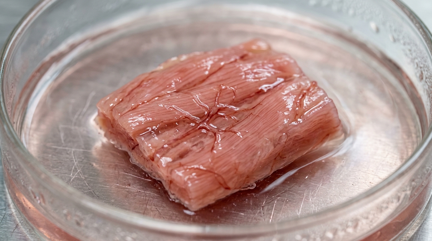
Engineers have taken a step toward laboratory-grown heart tissue that does more than just survive: it beats together. In a study published in the Journal of Pharmacy and Pharmaceutical Sciences, a research team at the University of Saskatchewan and the University of Manitoba used three-dimensional bioprinting to fabricate small cardiac patches seeded with human blood vessel cells, then showed that the patches with functioning vessel-like networks supported better organization and more synchronized beating of heart cells than patches without them. The work, led by Farinaz Ketabat and colleagues, addresses one of the most stubborn obstacles in cardiac tissue engineering: keeping thick, cell-dense constructs alive and functional by supplying them with an internal blood supply.
The problem the researchers set out to solve is well known in the field. Heart muscle depends on a dense network of capillaries to deliver oxygen and nutrients, and engineered cardiac tissue is no exception. Without vessels threading through a construct, cells deep inside can starve, and dead zones can form. Earlier work by the same group had shown that their alginate-gelatin bioink, a mixture of 3 percent alginic acid sodium salt and 1 percent gelatin, could support endothelial cell viability and print reliably. For the new study, they loaded that bioink with human umbilical vein endothelial cells, or HUVECs, at a density of 6 million cells per milliliter, and added two pro-angiogenic growth factors, vascular endothelial growth factor and basic fibroblast growth factor, each at 100 nanograms per milliliter, to encourage the embedded cells to form vessel-like structures.
Architecture mattered as much as chemistry. The team printed circular constructs 9 millimeters in diameter, built up in 16 layers, with each layer rotated 10 degrees relative to the one beneath it. The first layer ran at minus 70 degrees and the last at plus 80 degrees, a deliberate echo of the helical arrangement of muscle fibers in the native left ventricle, where fiber orientation sweeps from the outer epicardium to the inner endocardium and contributes to the twisting contraction that drives blood circulation. Printing proceeded at 4 millimeters per second under 25 kilopascals of pressure through a 27-gauge tapered tip, and the finished constructs were cross-linked in a calcium chloride solution, washed, and impregnated with type I collagen, which the authors note serves both as a structural scaffold for vessel formation and as a reservoir that modulates pro-angiogenic factors.
Once printed, the constructs were divided into three experimental groups. A control group was kept in static culture with no growth factors and no mechanical conditioning. A second group received the two growth factors during the first 10 days of culture but remained static. A third group received the growth factors and was then transferred on day 10, when endothelial network formation had begun, to a bioreactor that subjected the constructs to both compressive loading and fluid perfusion for one hour per day over three consecutive days. In the bioreactor, the constructs were compressed with 3 percent strain at a frequency of 1 hertz, matching the physiological rhythm of heart muscle, while medium was perfused through them at 5 milliliters per minute at body temperature. The authors chose these parameters to remain within the elastic region of the material, as determined in their previous work, and to partially simulate the mechanical and fluid environment of a beating heart.
The differences between the groups were visible under the microscope. Live-dead staining at the end of a 21-day culture period showed that control constructs had limited cell viability, with cells remaining mostly spherical and showing no endothelial organization. Constructs treated with growth factors alone fared better, displaying increased viability and early signs of endothelial organization, but the cells were distributed randomly and network formation was only partial. It was the combination of growth factors and bioreactor conditioning that produced the clearest result: markedly enhanced viability and the formation of continuous, interconnected endothelial networks throughout the constructs. Immunofluorescence confirmed that these networks expressed CD31, an endothelial marker, along continuous vessel-like structures, and also showed connexin 43, a gap junction protein, suggesting intercellular communication among the vessel cells. A quantitative check on four sectioned vascularized constructs found a CD31-to-DAPI fluorescence ratio of 0.98 plus or minus 0.03, indicating that endothelial cell distribution was reproducible from construct to construct.
With vascularized and non-vascularized constructs in hand, the researchers seeded heart cells onto them and asked whether the preformed vessel networks changed how the cardiac cells behaved. They used three cell types in sequence. H9c2 cells, a widely used rat-derived cardiac cell line, served for preliminary studies; these cells adhered and expressed the structural protein ACTN2 but did not contract, and showed no distinct sarcomeric organization, indicating they remained immature in this setting. The more demanding tests used neonatal rat ventricular myocytes, freshly isolated from two-day-old Sprague-Dawley rat pups, and human induced pluripotent stem cell-derived cardiomyocytes, both seeded at 500,000 cells per construct after roughly 15 to 16 days of pre-culture.
Structural staining told a consistent story across both primary cardiac models. Neonatal rat ventricular myocytes seeded onto non-vascularized constructs expressed cardiac troponin T and showed typical morphology, but had no clear association with any endothelial structures. On vascularized constructs, the same cells not only expressed troponin T but preferentially adhered to and aligned with the CD31-positive networks, integrating with the preformed vessel-like architecture. Semi-quantitative analysis of representative imaging fields, which the authors explicitly describe as limited by small numbers of biological replicates, estimated troponin T-positive coverage at 43.6 percent on vascularized constructs versus 31.5 percent on non-vascularized ones. The human cells showed an even starker contrast: induced pluripotent stem cell-derived cardiomyocytes on non-vascularized constructs formed rounded, compact clusters with limited spreading, while on vascularized constructs they spread into elongated, aligned, continuous troponin T-positive regions spatially associated with the endothelial networks, with estimated coverage of 52.6 percent versus 23.4 percent.
Function tracked with structure. Using calcium imaging with the fluorescent dye Fluo-8, the team recorded spontaneous activity in the seeded cells. One day after seeding, neonatal rat myocytes on non-vascularized constructs were rounded and isolated but beat spontaneously; on vascularized constructs they were more elongated and aligned, and their contractions were synchronized and more rhythmic. By day 21, cells on non-vascularized constructs still contracted but irregularly and arrhythmically, whereas those on vascularized constructs retained coordinated, synchronous beating with a more interconnected cellular network, though at a slightly reduced beating frequency. The human cardiomyocytes showed the same pattern at day 21: on non-vascularized constructs, beating occurred in isolated clusters that required separate analysis regions, with more frequent but unsynchronized contractions, while on vascularized constructs the cells formed continuous aligned networks that contracted synchronously across the entire field of view, again at a lower spontaneous frequency. The authors interpret the lower frequency alongside the higher synchronization as evidence of improved functional integration rather than impaired activity.
The authors connect these observations to known biology. Calcium transients reflect excitation-contraction coupling, the process linking electrical activation to mechanical contraction, so more regular transients suggest more coordinated function. They also cite prior work indicating that endothelial cells do more than deliver nutrients: they produce cardioactive factors in response to substances released during perfusion, and these factors can influence both the strength and duration of contraction in neighboring cardiomyocytes. Consistent with that idea, the presence of vascular cells has been shown in other studies to enhance maturation and contraction force in human cardiac organoids, possibly by keeping cells within a minimal diffusion distance of oxygen and nutrients. The Saskatchewan team notes that their combination of a thick, 16-layer bioprinted architecture mimicking myocardial alignment with both perfusion and mechanical stimulation distinguishes their approach from earlier studies that paired VEGF and bFGF with flow alone.
The study has clear limits, which the authors state directly. The culture period was only 21 days, so longer-term maturation and degradation of the hydrogel remain unexplored. The calcium analysis was qualitative and semi-quantitative, without measurements of transient kinetics such as rise time, decay time, or duration, and without force generation measurements that would directly quantify contractile strength. Bioreactor capacity, four constructs per run, and the scarcity of primary cardiac cells constrained the experimental design, forcing a choice between whole-mount and sectioned imaging for different samples. The authors also flag that future work should optimize the timing of cardiomyocyte seeding and the bioreactor conditions. Even so, the demonstration that a printed, helically layered scaffold can host both an endothelial network and synchronously beating cardiac cells, in both a pre-clinical rodent model and a human cell model, offers a concrete template for the next generation of engineered heart tissue, one in which the plumbing and the pumping are designed together from the first printed layer.
Subject of Research: Three-dimensional bioprinting of vascularized cardiac tissue constructs that support improved cardiomyocyte organization and synchronized contractile behavior
Article Title: 3D bioprinting of vascularized cardiac constructs reveals improved cardiomyocyte organization and contractile function
Article References: Ketabat, F., Kim, A., Alizadeh Sardroud, H., Eames, B. F., Collins, M., Chen, X., Kelly, M. E., & Badea, I. (2026). 3D bioprinting of vascularized cardiac constructs reveals improved cardiomyocyte organization and contractile function. Journal of Pharmacy & Pharmaceutical Sciences, 29, Article 17204. https://doi.org/10.3389/jpps.2026.17204
Image Credits: AI Generated
Keywords: 3D bioprinting, cardiac tissue engineering, vascularization, endothelial cells, cardiomyocytes, bioreactor, calcium transients, HUVECs, alginate-gelatin bioink, left ventricle, VEGF, bFGF
News Source: Gregory Coleman. (November 6, 2026). Bioprinted Heart Tissue With Built-In Blood Vessels Beats in Sync, Study Finds. Scienmag.