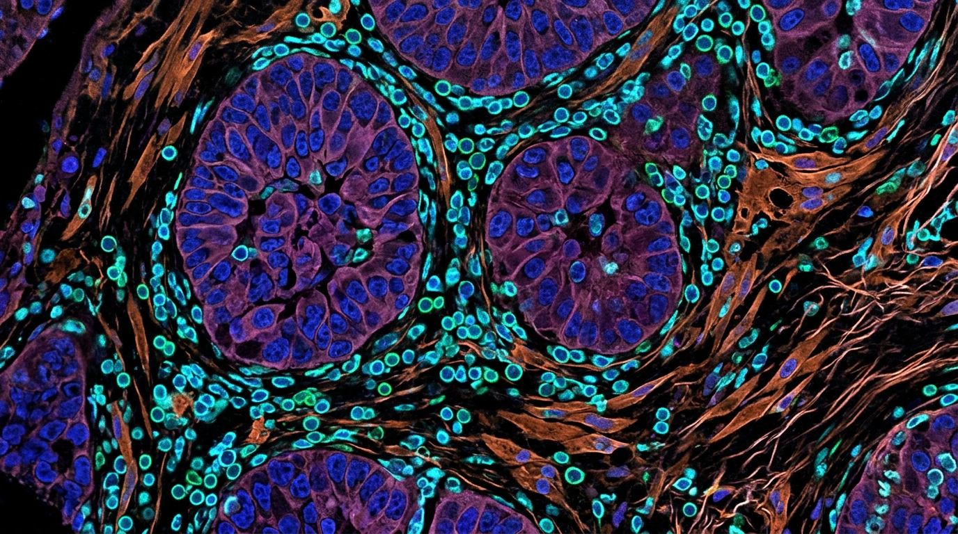
The tumor microenvironment functions as a complex, multicellular ecosystem that fundamentally shapes tumor initiation, progression, and therapeutic responses. Comprising immune and stromal compartments alongside vascular and neural-glial components, this environment coevolves with malignant cells to govern the success or failure of antitumor immunity. Recent technological breakthroughs, specifically high-resolution single-cell and spatial transcriptomic atlases, have moved the field beyond simple cell-type inventories toward a comprehensive understanding of the tumor microenvironment as a system of coordinated cellular networks. By cataloging the full cellular repertoire and functional states within tumors, researchers have identified tumor-enriched, specialized cell populations such as CXCL13+ T cells, SPP1+ tumor-associated macrophages, LAMP3+ dendritic cells, and LRRC15+ cancer-associated fibroblasts. These subsets do not act in isolation but assemble into functional modules and immunity hubs that dictate the spatial organization and immune-reactive or immune-evasive configurations of solid tumors.
T cells remain the central effectors of antitumor immunity, with CD8+ cytotoxic T lymphocytes playing the primary role in recognizing tumor antigens and directly killing malignant cells. Within the immunosuppressive tumor microenvironment, however, these cells often transition into functionally exhausted states characterized by diminished effector activity and sustained inhibitory receptor expression. Single-cell analyses across multiple cancer types have consistently identified these exhausted populations, while also uncovering CXCL13+ pre-exhausted CD8+ T cells that retain antitumor potential and correlate with improved responses to immune checkpoint blockade. CD4+ T cells exhibit even greater plasticity, differentiating into various effector subsets that can either support or suppress cytotoxic responses. CXCL13+ Th1-like CD4+ T cells, in particular, have emerged as a critical tumor-reactive population that recruits B cells and promotes the formation of tertiary lymphoid structures, thereby amplifying local immune responses. Conversely, TNFRSF9+ activated regulatory T cells maintain an immunosuppressive balance, and their accumulation is a hallmark of poor clinical outcomes in several cancers, providing a clear target for therapeutic strategies designed to alleviate immune suppression.
B cells have transitioned from passive bystanders to central modulators of antitumor immunity, demonstrating remarkable phenotypic and functional heterogeneity. Pan-cancer single-cell analyses have delineated two primary differentiation programs: an organized germinal-center-like pathway associated with superior survival and strong immunotherapy responses, and an extrafollicular pathway that is often linked to immune resistance and a worse prognosis. In the context of tertiary lymphoid structures, which resemble secondary lymphoid follicles and are associated with improved prognosis, memory B cells and plasma cells contribute to tumor-specific humoral responses. While certain regulatory B-cell subsets secrete immunosuppressive cytokines like IL-10 to attenuate cytotoxic responses, the overall functional balance between effector and regulatory B cells often dictates the net impact on tumor growth. Consequently, the composition of the intratumoral B-cell compartment is becoming an important determinant for patient stratification and immunotherapy efficacy, as researchers look for ways to augment beneficial humoral functions while suppressing deleterious regulatory subsets.
Myeloid cells, particularly tumor-associated macrophages and dendritic cells, serve as the innate foundation of the tumor microenvironment. SPP1+ tumor-associated macrophages have been identified as a pro-angiogenic population linked to poor clinical outcomes across various cancer types, contrasting with inflammatory macrophage subsets that co-localize with activated CD8+ T cells in immune hubs. Dendritic cells, specifically the mature LAMP3+ subset, are critical for initiating and coordinating robust adaptive immune responses through their ability to migrate from tumors to lymph nodes. Recent studies show that cDC1-derived LAMP3+ dendritic cells exhibit preferred antitumor functions and correlate with higher response rates to immune checkpoint blockade, while their depletion during tumor progression impairs the priming of tumor-specific T cells. Pharmacological strategies, such as the use of phosphodiesterase-5 inhibitors, show promise in reversing homing defects and restoring the migratory capacity of these cells to rejuvenate antitumor immunity in late-stage tumors.
Cancer-associated fibroblasts represent a highly heterogeneous component of the tumor stroma, with distinct transcriptional states forming a continuum from tumor-restraining to tumor-promoting phenotypes. LRRC15+ myofibroblasts have been identified as a terminal pro-tumorigenic state associated with immune-excluded microenvironments, where they actively dampen the infiltration and activation of CD8+ T cells. Other subsets, such as the PDGFRα+ITGA11+ lymphangiogenic fibroblasts and metabolic fibroblasts driven by nicotinamide N-methyltransferase, further remodel the extracellular matrix and provide supportive niches for immune evasion. The presence of dense, collagen-rich myofibroblasts at the tumor periphery is frequently linked to poor patient survival, underscoring the need to target specific pro-tumorigenic fibroblast programs rather than treating the stromal compartment as a homogeneous entity. By identifying these state-specific markers, researchers aim to develop therapies that reprogram the stroma toward a more homeostatic, immune-permissive state.
Tumor vasculature is no longer viewed as a passive conduit but as a dynamic immunological hub that can either facilitate immune cell trafficking or contribute to immune exclusion. Single-cell mapping of tumor vasculature has revealed functional divergences between angiogenic tip cells, which correlate with aggressive tumor behavior and resistance to anti-angiogenic therapies, and SELE+ venous endothelial cells that facilitate proinflammatory signatures for leukocyte infiltration. Specialized structures like tumor-associated high endothelial venules represent key portals for T- and B-cell entry, and their maturation is often coupled to improved therapeutic responsiveness. Furthermore, therapy-induced endothelial reprogramming, such as the emergence of ACKR1+ endothelial subsets, creates positive feedback loops with effector T cells, converting the vascular landscape into a scaffold for recruitment and sustainment of antitumor immunity. This refined portrait suggests that combining vascular normalization strategies with immunotherapy may synergistically improve drug delivery and immune cell infiltration.
The field of cancer neuroscience has brought attention to the complex neural-glial components within the tumor microenvironment that regulate tumor growth, metastasis, and immune responses. Peripheral nerves infiltrating tumors release neurotransmitters and neurotrophic factors that not only fuel cancer cell proliferation but also reshape the immune landscape into a tumor-permissive state. Schwann cells have been found to guide migrating cancer cells through the tissue, forming physical tracks that facilitate perineural spread. Spatial profiling has further elucidated an immunosuppressive versus immunopermissive dichotomy, where nerve infiltration by tumor cells fosters an immune-cold niche characterized by proinflammatory fibroblasts and exhausted T cells, whereas non-invaded nerves support immune surveillance. These inter-organ neuroimmune circuits provide a basis for repurposing neuroactive medications, such as antiepileptics or beta-blockers, to attenuate nerve-mediated tumor support and enhance the efficacy of conventional anticancer regimens.
Beyond characterizing individual cell types, the current research trend is shifting toward an integrated view of multicellular coordination, defined as cellular modules or immunity hubs. These spatially and functionally organized units, containing coordinated clusters of T cells, dendritic cells, and stromal components, function as integrated ecosystems that drive immune surveillance or disease progression. Data from pan-cancer atlases suggest a common model of tumor evolution, characterized by the progressive disintegration of healthy, tissue-specific multicellular networks and the gain of highly conserved cancer-associated modules. Understanding how these immune and stromal cell subsets cooperate as a system is essential for developing broad-spectrum, tumor-microenvironment-targeted therapies that address the higher-order interactions governing resistance to current treatments.
Artificial intelligence is becoming an indispensable tool for synthesizing this large-scale, complex data, enabling the development of predictive models that go beyond static measurements. The concept of the AI-driven virtual cell leverages multimodal datasets and large language models to simulate cellular behavior and responses to perturbations, overcoming the limitations of traditional descriptive models. Looking ahead, this approach is being extended to the AI-driven virtual tumor, which aims to integrate cell states, spatial organization, and cell-cell communication networks into a unified, perturbable digital representation of the tumor ecosystem. By learning predictive rules from atlas data and clinical outcomes, these frameworks could eventually enable in silico hypothesis testing, refined patient stratification, and the rational design of network-informed combination therapies.
Despite these conceptual and computational advances, several methodological challenges remain in translating atlas-level insights into routine clinical practice. The field continues to grapple with data heterogeneity across platforms, batch effects, and the lack of standardized benchmarks for dynamic tumor microenvironment behaviors. Future progress will require the construction of high-quality, longitudinal, and multimodal atlases that capture the evolutionary dynamics of tumors under treatment, alongside rigorous wet-lab validation using patient-derived organoids and humanized mouse models. By moving from correlative observations to causal mechanisms, researchers hope to reshape the tumor immune ecosystem at the macroscopic, mesoscopic, and microscopic levels, ultimately delivering durable and precise clinical outcomes for a broader spectrum of patients.
Subject of Research: Systematic characterization of the tumor microenvironment using single-cell and spatial omics to inform precision cancer immunotherapy.
Article Title: The cellular actors of the tumor microenvironment: a single-cell atlas perspective on specialized subtypes, coordinated networks, and immunotherapy
Article References: Zhu, L., Jiang, N., & Zhang, Z. (2026). The cellular actors of the tumor microenvironment: a single-cell atlas perspective on specialized subtypes, coordinated networks, and immunotherapy. Immunity & Inflammation, 2(1), Article 27. https://doi.org/10.1007/s44466-026-00043-3
Image Credits: AI Generated
DOI: 10.1007/s44466-026-00043-3
Keywords: tumor microenvironment, single-cell sequencing, spatial transcriptomics, cancer immunotherapy, immune checkpoint blockade, cellular networks, T cells, cancer-associated fibroblasts, artificial intelligence, precision medicine, tumor-associated macrophages, tumor-infiltrating lymphocytes, tertiary lymphoid structures, computational modeling, virtual tumor models
News Source: Nathaniel Bowman. (November 18, 2026). Mapping Tumor Microenvironments Reveals Coordinated Networks and Guides Precision Immunotherapy. Scienmag.