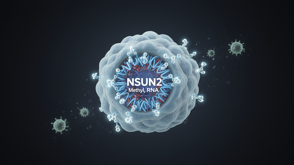
Lung adenocarcinoma is the most common and aggressive form of lung cancer, and although immune checkpoint inhibitors have reshaped its treatment, only roughly 20% of patients with this disease benefit from the drugs. A new study published in Experimental Hematology & Oncology points to a reason why, tracing that resistance to a single RNA-modifying enzyme inside tumor cells. The researchers report that the 5-methylcytosine (m5C) methyltransferase NSUN2 acts as a master switch for immune exclusion: high levels of the protein in tumor cells suppress the recruitment of cytotoxic CD8+ T cells, while deleting or inhibiting NSUN2 converts immunologically “cold” tumors into “hot” ones and markedly improves responses to anti-PD-1 therapy in mouse models.
Analyzing a public single-cell RNA sequencing dataset from non-small cell lung cancer patients treated with anti-PD-1 therapy, they identified 16 RNA methylation regulatory genes that were significantly upregulated in malignant cells of patients with stable disease compared with those achieving a partial response. Malignant cells from patients with stable disease expressed 2.4-fold more NSUN2 than those from responders. Cross-referencing this list with proteomic data from two independent lung adenocarcinoma cohorts narrowed the field to three candidates upregulated in tumors, with NSUN2 showing the most significant difference.
Independent datasets reinforced the association. In two additional bulk RNA sequencing cohorts of immune checkpoint blockade-treated patients, NSUN2 expression was consistently higher in non-responders than in responders. The Cancer Genome Atlas lung adenocarcinoma cohort and the CPTAC proteomic repository both showed significant NSUN2 elevation in tumors relative to normal tissue, and single-cell datasets indicated the overexpression was confined predominantly to tumor cells rather than other cell types in the microenvironment. High NSUN2 expression correlated with shorter overall survival in TCGA and across multiple lung cancer cohorts, and in a tissue microarray of 52 paired tumor and adjacent normal specimens, immunohistochemistry confirmed protein upregulation in tumors and significantly poorer overall survival among patients whose tumors scored high for NSUN2.
To test whether the enzyme mattered functionally, the researchers built several experimental systems: Lewis lung carcinoma cells with stable Nsun2 knockdown, KRAS/p53-mutant KP cells with complete CRISPR-Cas9 knockout, and a genetically engineered mouse model in which Nsun2 could be deleted specifically in the alveolar epithelial cells that give rise to this cancer subtype. In culture, NSUN2-deficient cells proliferated more slowly. In immunocompetent mice, however, the effect was far more dramatic: subcutaneous tumors lacking Nsun2 grew poorly, and spontaneous lung tumors in the conditional knockout mice showed significantly reduced tumor burden and longer overall survival. Strikingly, when the same tumor cells were implanted into immunodeficient Rag1-deficient mice, the growth inhibition largely disappeared, showing that NSUN2’s pro-tumor role in vivo depends mainly on the immune system rather than on cell-autonomous growth control.
Sequencing revealed what that immune dependence meant. Single-cell RNA sequencing of spontaneous tumors showed a 32% increase in total T and natural killer cell infiltration after Nsun2 loss, with CD8+ T cells rising from 35.8% to 62.3% of the relevant compartment while myeloid-derived suppressor cells fell from 64.7% to 35.3%. Flow cytometry confirmed more tumor-infiltrating CD8+ T cells, including more IFN-γ-producing and granzyme B-producing effector subsets, and immunofluorescence imaging showed denser CD8+ T cell penetration of the tumor parenchyma. One caveat emerged: terminally exhausted PD-1-positive TIM-3-positive CD8+ T cells also increased, which the authors interpret as a byproduct of sustained T cell activation rather than immune suppression, noting that the preserved synergy with anti-PD-1 suggests these cells remain partially responsive to checkpoint blockade.
The mechanistic thread connecting NSUN2 to T cell exclusion turned out to be the chemokine CCL5, which attracts CD8+ T cells expressing its receptor CCR5. After Nsun2 depletion, chemotaxis pathways were the most enriched gene sets in bulk RNA sequencing, and Ccl5 emerged as upregulated in malignant cells, showing 3.7-fold higher expression in knockout tumor cells by single-cell analysis. The increase held at the protein level in cell supernatants and in tumor interstitial fluid, and supernatants from Nsun2-deficient cells recruited significantly more primary CD8+ T cells in Transwell migration assays, including human CD8+ T cells migrating toward media from NSUN2-knockdown human lung cancer cells. In tumors, the CCR5-positive CD8+ T cell subset expanded consistently after Nsun2 depletion.
The pathway ran through an unexpected intermediary. m5C methylated RNA immunoprecipitation sequencing showed no change in the m5C status of Ccl5 transcripts themselves, but revealed significant hypomethylation of histone-modifying enzyme mRNAs after Nsun2 knockdown. Among twelve candidates whose transcripts lost m5C marks without changes in abundance, only Hdac8 knockdown robustly reproduced the Ccl5 upregulation, producing a 1.5-fold increase. Hdac8 mRNA showed the most dramatic methylation loss, and although its transcript levels were unchanged, ribosome nascent-chain complex qPCR demonstrated that its translational efficiency dropped after Nsun2 depletion. A dual-luciferase reporter carrying the Hdac8 5′ untranslated region and adjacent coding sequence confirmed the mechanism: NSUN2 overexpression enhanced translation of the wild-type reporter, but the enhancement vanished when the four identified m5C sites were mutated.
The reader protein completing the cascade was YBX1. RNA immunoprecipitation showed Hdac8 mRNA robustly pulled down by anti-YBX1 antibody in control cells but not after Nsun2 depletion, while two other candidate readers showed no enrichment. Knocking down Ybx1 reduced HDAC8 protein without touching its mRNA and consequently raised Ccl5 expression. Downstream, HDAC8 repressed CCL5 epigenetically: the selective HDAC8 inhibitor PCI-34,051 increased Ccl5 mRNA more than 2-fold and secreted protein more than 90%, raised global H3K27 acetylation, and enhanced H3K27ac occupancy at the Ccl5 promoter as measured by ChIP-qPCR. In rescue experiments, an HDAC8 inhibitor reversed the CCL5 suppression and impaired CD8+ T cell migration caused by NSUN2 overexpression, establishing NSUN2-m5C-YBX1-HDAC8-CCL5 as a coherent regulatory axis.
Clinical specimens supported the translational relevance. In a multiplex immunohistochemistry analysis of 43 primary lung adenocarcinoma samples, NSUN2-high tumors showed sparse CD8+ T cell infiltration while NSUN2-low tumors were densely infiltrated, yielding a statistically significant inverse correlation. In a separate cohort of 18 patients who received neoadjuvant anti-PD-1 or anti-PD-L1 therapy before surgery, those achieving pathological complete or major partial responses had significantly lower intratumoral NSUN2 and higher CD8+ T cell infiltration than pathological non-responders. Depletion experiments in mice confirmed causality in the other direction: anti-CD8 antibody treatment abolished the anti-tumor benefit of Nsun2 knockout, while MDSC depletion and Treg ablation failed to do so, indicating those myeloid and regulatory changes were secondary consequences.
Therapeutically, combining Nsun2 genetic depletion with anti-PD-1 produced synergistic tumor regression in KP and LLC allograft models, with tumor remission exceeding 80% in the combination groups, and the dual strategy also significantly reduced tumor burden in the spontaneous model. Neutralizing CCL5 with an antibody reversed the growth inhibition conferred by Nsun2 depletion, cementing the chemokine as the required mediator. Pharmacologically, the team encapsulated the small-molecule NSUN2 inhibitor MY-1B in iRGD-functionalized liposomes, nanocarriers whose surface peptide homes to tumor vasculature and drives transport into tumor tissue. The liposomal formulation showed no significant toxicity in tumor-naïve mice and, combined with anti-PD-1, produced robust anti-tumor responses in both allograft models. The authors acknowledge limitations: the efficacy studies used subcutaneous rather than orthotopic models, the immunotherapy-treated clinical cohort was small, and the full spectrum of NSUN2’s m5C targets in immunity remains to be mapped. Within those bounds, the work positions NSUN2 as both a candidate biomarker for identifying patients unlikely to benefit from checkpoint inhibitors and a druggable node for converting cold lung tumors into ones that immunotherapy can reach.
Subject of Research: The role of the m5C methyltransferase NSUN2 in suppressing CD8+ T cell infiltration and immunotherapy response in lung adenocarcinoma
Article Title: Tumor-intrinsic NSUN2 orchestrates immunosuppression in lung adenocarcinoma via the m5C-HDAC8-CCL5 axis
Article References: Meng, W., Lu, T., Du, M., Dai, X., Han, Y., Li, Y., Xu, H., Yang, B., Wang, Z., Du, H., Liao, K., Liu, X., Sun, Y., Yang, C., Dong, D., Yan, Y., Guo, W., Li, B., & Li, H. (2026). Tumor-intrinsic NSUN2 orchestrates immunosuppression in lung adenocarcinoma via the m5C-HDAC8-CCL5 axis. Experimental Hematology & Oncology, 15(1), Article 89. https://doi.org/10.1186/s40164-026-00826-9
Image Credits: AI Generated
DOI: 10.1186/s40164-026-00826-9
Keywords: NSUN2, lung adenocarcinoma, m5C methylation, HDAC8, CCL5, CD8+ T cells, immune checkpoint inhibitors, YBX1, tumor microenvironment, anti-PD-1 therapy, immunotherapy resistance, iRGD liposomes
News Source: Nathaniel Bowman. (October 29, 2026). RNA Methylation Enzyme NSUN2 Keeps Lung Tumors Cold and Blocks Immunotherapy. Scienmag.