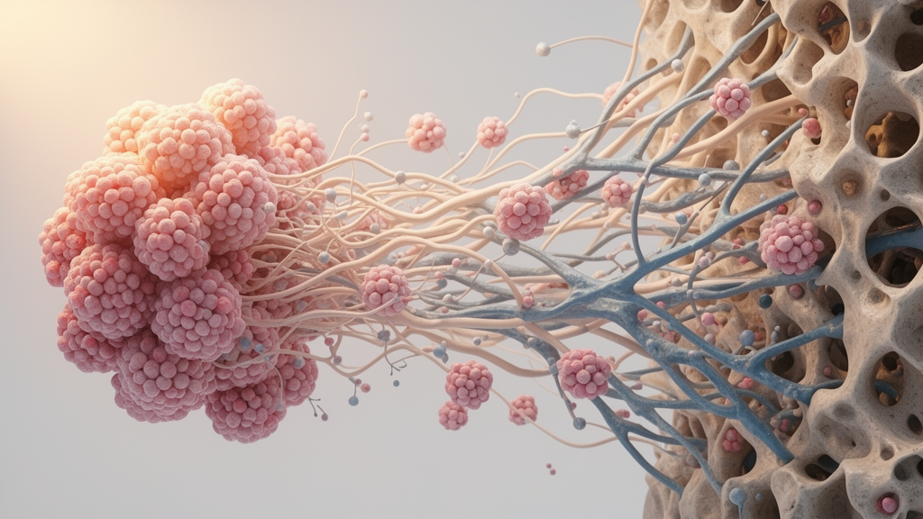
Osteosarcoma cells may survive treatment and spread to the lung partly by changing how they interact with the extracellular matrix, according to a 2026 focused narrative Review. The authors propose “matrix-state switching” as a stage-resolved framework in which ligand presentation, adhesion-receptor usage, matrix-coupled signaling and the participating cellular compartment jointly determine tumor behavior. The available evidence supports different functional states in the primary tumor, during detachment and after pulmonary re-adhesion, but it does not yet prove that the same clone makes each transition. Observed differences could instead reflect reversible adaptation, selection of pre-existing subpopulations, treatment-associated remodeling or a combination of these processes.
The Review divides the evidence into three selective contexts. State I comprises attached cells growing in the mineralized, collagen-rich and fibronectin-rich primary environment; State II captures the detachment bottleneck, when conventional focal-adhesion signaling is disrupted and survival requires tolerance of anchorage loss; State III represents pulmonary re-adhesion and niche construction. This division is not a compulsory metastatic sequence. Cells may enter dormancy, circulate as matrix-bearing clusters, obtain platelet or fibrin shielding, or reach the lung after selection of dissemination-competent subclones. The framework therefore asks which receptor–ligand–signaling architecture is necessary in a particular context, rather than assuming that repeated AKT activation reflects one continuous ECM–integrin–FAK pathway.
Primary-tumor evidence indicates that matrix protection is component-selective rather than universal. In a patient-derived osteosarcoma culture model, cells received a four-hour pulse of methotrexate, cisplatin, maphosphamide or doxorubicin before being placed beneath an extracellular-matrix gel. Apparent half-maximal inhibitory concentrations were higher after matrix transfer for all four agents, with the largest difference after doxorubicin, although numerical values were not reported. On plastic, doxorubicin produced extensive DNA fragmentation, while matrix-exposed cells showed less damage, reduced total p53 and prevented efficient nuclear p53 accumulation. Heparan sulfate proteoglycan provided the strongest protection and fibronectin a weaker effect; laminin and nidogen did not reduce doxorubicin cytoreduction, whereas collagen IV increased it. Because p53 re-expression or nuclear-targeting rescue was not performed, altered repair, cell-cycle behavior or apoptosis thresholds remain possible explanations.
Other studies resolve distinct modules rather than one universal resistance mechanism. TGFBI, also called βig-h3, bound α2 and β1 integrin subunits and connected them to PI3K–AKT signaling: depletion reduced adhesion, invasion and migration in SaOS-2 and MG-63 cells, while receptor blockade reproduced the effect. Separately, matrix-associated TIMP1 was associated with surface αvβ3 and anti-apoptotic activity under a TNF-α plus cycloheximide model; disrupting αvβ3 ligation reduced the effect of recombinant TIMP1 by about 30%. These experiments do not establish the same survival architecture. Their models also differ in treatment and context, reinforcing the need to measure ligand identity, receptor state and exposure sequence within the same experimental window. In clinical material, a retrospective series of 36 high-grade osteosarcomas found higher FAK expression in poor histologic responders, and an internally derived β5–FAK–GSK3β profile achieved an area under the curve of 0.899. Its 94% sensitivity, 86% specificity and 90% accuracy remain hypothesis-generating because the cohort was small and the model lacked external validation.
Detachment exposes the limitations of a single focal-adhesion model. In parental SAOS-2 cells, suspension increased apoptosis from 10.3% to 51.8% and triggered mitochondrial cytochrome-c release and caspase-3 activation. Repeated suspension selection produced anoikis-resistant cells in which Src and AKT remained active; inhibiting Src or PI3K restored anoikis even though FAK and paxillin were dephosphorylated, and constitutively active FAK did not rescue the sensitive cells. This defines a FAK-independent Src–PI3K–AKT survival topology in that selected model. It should not be generalized to every osteosarcoma or assumed to predict behavior after re-adhesion. Other work has linked FASN–ERK/Bcl-xL, ID1–PI3K–AKT and FOSL1–SOX2 programs to anoikis resistance or metastatic fitness, illustrating a network of parallel, non-equivalent adaptations.
Loss of static adhesion can even coexist with greater metastatic fitness. In U-2 OS cells, reducing caveolin-1 lowered adhesion to collagen I, collagen IV, fibronectin and laminin by 50%–80%, yet increased soft-agar growth, survival on poly-HEMA, migration, invasion and experimental lung colonization. Mice receiving caveolin-1-deficient cells developed more than 250 lung metastases, compared with a median of two after parental cells and none after caveolin-1-overexpressing cells. The deficient cells showed greater c-Src abundance and activity, and a Src inhibitor suppressed their migration. These findings make clear that receptor abundance alone cannot predict function: integrin conformation, ligand occupancy, clustering, adaptor recruitment, mechanical context and survival signaling can all change together. Accordingly, the detached phase should be treated as a transient bottleneck that may offer a distinct treatment window rather than as a uniformly weaker version of attached growth.
Pulmonary colonization introduces another selective bottleneck because surviving cells must regain adhesive competence in a matrix and stromal environment unlike that of the primary bone lesion. Historical paired pathology showed that every evaluable pulmonary metastasis differed from its paired primary lesion in at least part of the integrin repertoire. A functional example is β4 integrin in highly metastatic MNNG-HOS cells: knockdown did not materially alter monolayer proliferation and had inconsistent effects in routine migration or invasion assays, yet markedly reduced anchorage-independent growth and experimental lung colonization. Disruption of β4 Tyr1494 reduced AKT and S6 phosphorylation, lung metastasis and mortality while affecting primary intramuscular growth much less. β4 associated with ezrin, and β4 expression re-emerged in late lesions, consistent with selection for escape variants or receptor recovery. This module concerns pulmonary colonization, but the tail-vein model bypassed early dissemination and did not establish a linear β4-to-ezrin pathway or a laminin ligand.
The lung niche can also be constructed by stromal cells rather than exclusively through tumor-cell-autonomous receptor switching. Single-cell profiling, fibroblast assays, mouse experiments and a patient-derived xenograft model identified a CXCL14-high osteosarcoma population with stem-like and anoikis-resistant properties. Tumor-derived CXCL14 engaged α11β1 on fibroblasts, activating AKT and ERK signaling, focal-adhesion maturation, actomyosin contractility and matrix contraction. The stimulated fibroblasts produced TGF-β, and the circuit was associated with exclusion of CD8-positive T cells. Blocking the CXCL14–α11β1 axis reduced fibroblast TGF-β production, improved T-cell activity and suppressed lung metastasis. Because α11β1 was principally a fibroblast receptor in this model, bulk ITGA11 or ITGB1 expression cannot be interpreted as evidence of a malignant-cell receptor state. The findings demonstrate a tumor–stroma niche mechanism, although they do not establish resistance to a particular osteosarcoma immunotherapy regimen or direct interaction with the β4 pathway.
Recent paired analyses strengthen the case for therapy-associated ecosystem remodeling while preserving the distinction between association and causation. A matched study of three patients before and after neoadjuvant chemotherapy found expanded fibroblast and endothelial compartments, contracted immune populations, and residual malignant-cell programs involving epithelial–mesenchymal transition, extracellular-matrix interaction and integrin/PI3K-related signaling. The paired design improves temporal inference, but the small cohort, transcript-level measurements, pseudotime reconstruction and computational drug-sensitivity predictions cannot distinguish treatment-induced changes from selective enrichment of pre-existing states. The Review therefore recommends longitudinal lineage tracing, spatial protein validation and perturbation. Clinically, the most informative series would pair pretreatment biopsy, post-chemotherapy resection, pulmonary metastasis and recurrent tissue, looking for changes in matrix modules rather than relying on the absolute abundance of a single marker in bulk tissue.
LAMC1 illustrates how easily a transcriptomic candidate can be mistaken for an established mechanism. Osteosarcoma-specific evidence currently includes heterogeneous LAMC1 messenger RNA expression among cell lines and a TARGET-derived survival association, but no demonstration of γ1 protein abundance, laminin heterotrimer assembly, extracellular deposition, receptor engagement, state-specific signaling or a perturbation-and-rescue phenotype. Historical pathology found weak or absent laminin staining in most primary tumors, creating a testable rather than contradictory possibility: a laminin dependency might be absent from much of the primary lesion but emerge in a pulmonary matrix. A functional test would require LAMC1 depletion, confirmation of the assembled γ1-containing laminin in the relevant compartment, identification of its receptor, a state-specific phenotype and rescue by LAMC1 re-expression or the appropriate laminin matrix. Failure at those steps would appropriately leave LAMC1 as a correlated marker rather than a functional dependency.
The Review proposes a linked experimental sequence in which the same population moves from a defined primary-like matrix, through poly-HEMA or ultra-low-attachment suspension, to a defined lung-like matrix with or without fibroblasts. Each transition would be assessed for ligand assembly and deposition, receptor abundance and activation, FAK, Src and PI3K–AKT signaling, anoikis, re-adhesion, invasion, drug response and metastatic outgrowth. Genetic barcoding or lineage tracing could distinguish adaptation from clonal selection, while defined hydrogels, tissue-derived matrices, spheroids and organ-on-chip platforms offer different levels of control and realism. The Review’s central implication is not that clinicians should immediately target one extracellular-matrix pathway. Rather, experiments should identify the nonredundant dependencies that allow cells to move between survival states, potentially revealing time-limited vulnerabilities that stable monolayer assays cannot detect.
Subject of Research: Stage-specific extracellular-matrix and integrin signaling in osteosarcoma treatment resistance and pulmonary metastasis
Article Title: Matrix-state switching in osteosarcoma under therapeutic pressure: integrin rewiring, residual disease, and pulmonary niche adaptation
Article References: Gu, Y., Zhuang, Y., & Zhang, W. (2026). Matrix-state switching in osteosarcoma under therapeutic pressure: integrin rewiring, residual disease, and pulmonary niche adaptation. Frontiers in Cell and Developmental Biology, 14, Article 1987843. https://doi.org/10.3389/fcell.2026.1987843
Image Credits: AI Generated
DOI: 10.3389/fcell.2026.1987843
Keywords: osteosarcoma, extracellular matrix, integrin rewiring, anoikis resistance, lung metastasis, tumor microenvironment, therapy resistance, LAMC1, patient-derived models, spatial transcriptomics, Matrix-state, switching
News Source: Nathaniel Bowman. (October 24, 2026). Osteosarcoma rewires matrix signaling as disease progresses. Scienmag.