Researchers in China have reported that a course of magnetic seizure therapy, an emerging convulsive treatment for severe treatment-resistant depression, was followed by substantial short-term reductions in depressive and anxiety symptoms in a small group of patients. The same study, however, found that a much-publicized candidate brain signal—a drop in task-related frontal theta activity measured by electroencephalography—did not hold up under statistical correction, and the authors themselves caution that the design cannot establish causal mechanisms or biomarker value. The work, published in Frontiers in Psychiatry, is an exploratory, single-arm, pre–post study rather than a controlled trial, and its conclusions are deliberately framed as hypothesis-generating.
Magnetic seizure therapy, or MST, was developed as a focal alternative to electroconvulsive therapy. Both treatments induce a therapeutic seizure under anesthesia, but MST does so with high-intensity transcranial magnetic stimulation rather than electrical current passed through the brain. Because the induced magnetic field is more spatially restricted, prior studies have reported faster reorientation after treatment and fewer cognitive side effects, particularly less disruption of autobiographical memory. Randomized evidence has suggested that MST may achieve antidepressant outcomes comparable to ultrabrief right unilateral ECT, and a systematic review of fifteen studies involving three hundred participants reported variable but clinically relevant response rates. What has remained unclear is how MST changes brain function at the level of cognitive-control circuits, which is the question the new study set out to probe.
The research team enrolled nineteen patients with treatment-resistant depression, defined as non-response to at least two adequate antidepressant trials from different pharmacological classes. All met DSM-5 criteria for major depressive disorder. The group was unusual in several respects: sixteen of the nineteen participants were female, the mean age was 20.89 years with a range from 13 to 40, and ten of the nineteen were minors, whose participation required written assent alongside parental informed consent.
Treatment followed a standardized protocol. MST was delivered with a magnetic stimulator using a double-cone coil positioned over the vertex, targeting the bilateral F3 and F4 frontal regions, at 100 Hz and full device output in ten-second trains. Sessions were given five times weekly, with each participant receiving between six and twelve treatments, averaging 8.2, with the number determined by clinical response and tolerability. Every session was performed under general anesthesia induced with propofol and muscle relaxation with succinylcholine, and seizure activity was monitored by EEG, with an adequate seizure defined as lasting longer than ten seconds. Psychotropic medications were held at stable doses for at least two weeks before baseline and throughout the course, so no medication changes occurred during treatment.
Clinical outcomes were striking in magnitude. On the 24-item Hamilton Depression Rating Scale, scores fell from a mean of 40.53 at baseline to 22.53 after treatment, a paired t-test yielding t(18) = 7.206, P < 0.001, with a Cohen’s d of 1.653. Montgomery-Åsberg scores dropped from 35.68 to 20.68 (d = 1.702), and Hamilton anxiety scores from 28.53 to 16.74 (d = 1.279). With response defined as at least a fifty percent reduction from baseline, response rates were 42.1 percent on the Hamilton scale, 42.1 percent on the anxiety scale, and 57.9 percent on the Montgomery-Åsberg scale. Remission, however, was rare: zero percent on the Hamilton scale, 5.3 percent on the anxiety scale, and 10.5 percent on the Montgomery-Åsberg scale. The authors attribute this gap to the severity of the baseline symptoms, the relatively short course of about eight sessions, and the timing of assessment within 24 to 48 hours after the final treatment, which captures acute improvement rather than sustained recovery.
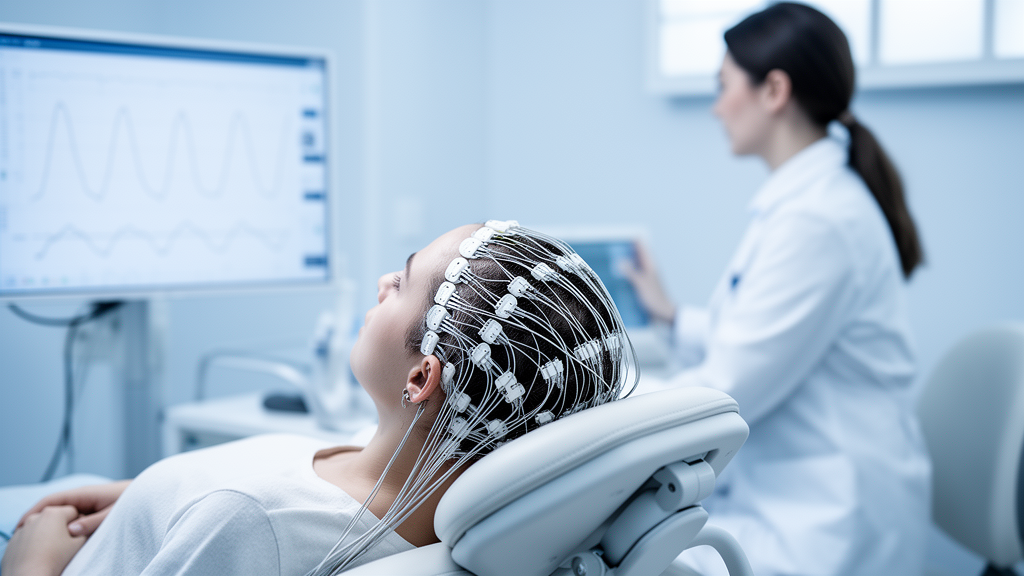
The mechanistic core of the study was a visual Go/Nogo task recorded with a 64-channel EEG system before and after the treatment course. In this task, participants respond quickly to one digit and withhold responses to another, with the mapping reversed between two blocks of 240 trials each. Response inhibition and conflict monitoring of this kind engage a network including the anterior cingulate cortex, dorsolateral prefrontal cortex, and inferior parietal regions. The researchers analyzed event-related potentials such as N2 and P3, time-frequency power in the theta and beta bands, EEG microstates, and theta–beta cross-frequency coupling between frontal and central electrodes. Frontal midline theta, typically 4 to 7 Hz, is linked to conflict detection and top-down control recruitment in anterior cingulate and dorsomedial prefrontal circuits, and in severe depression exaggerated task-related theta has been interpreted as inefficient or compensatory recruitment rather than effective inhibition.
The headline EEG result was nominal rather than confirmed. Total Nogo-related theta-band activity at the Fz electrode was lower after treatment, t(17) = −2.864, P = 0.011, d = −0.645, and the FCz electrode showed a similar nominal reduction, P = 0.044. But after Benjamini–Hochberg false discovery rate correction across all ten predefined time-frequency windows, neither effect survived, with corrected q values of 0.108 and 0.219. No comparable change appeared in the Go condition, yet because the study did not test a formal Time × Condition interaction, the authors state that condition specificity cannot be claimed. Conventional ERP amplitudes—N2, P3, and their difference waves—showed no significant changes at all, with effect sizes below 0.30, and theta–beta cross-frequency coupling during Nogo trials was unchanged.
The exploratory microstate analysis fared no better under correction. In the Go condition, Map4 area under the curve was nominally lower after treatment and its center of gravity shifted later; in the Nogo condition, Map2 duration was nominally longer. None of these survived false discovery rate correction, with q values ranging from 0.095 to 0.607. The authors are explicit that microstate maps are data-driven, their numbering is dataset-dependent, and functional labels such as attentional orienting or inhibitory preparation are tentative. They also note a technical caveat on the theta measure itself: because the 4–7 Hz analysis windows contained fewer than two full theta cycles, the findings should be read as changes in total theta-band activity rather than pure oscillatory amplitude, and the differing baseline windows used for ERP and time-frequency analyses may have contributed to the dissociation between the two measures.
A further blow to interpretation came from the behavioral data. Although reaction times, accuracy, and commission errors were recorded during the task, a technical storage error left the behavioral dataset incomplete for a subset of participants, precluding formal analysis. The EEG data itself remained intact—one participant’s recordings were corrupted and excluded, leaving eighteen for all EEG analyses—but without behavior the researchers cannot determine whether the nominal theta reduction was associated with improved inhibition, a speed–accuracy tradeoff, altered attention or engagement, or no behavioral change at all. They also emphasize that no normative comparison was performed, so whether baseline theta was abnormal or post-treatment theta approached normal levels cannot be known, and a reduction in frontal theta is not inherently beneficial since the signal can index effort, arousal, or task engagement as well as control.
The limitations section is unusually candid. As an open-label, single-arm, pre–post design, the study cannot attribute observed changes specifically to MST; practice effects, regression to the mean, symptom fluctuation, expectancy, anesthesia, and nonspecific seizure-related effects all remain possible. Residual postictal EEG slowing at the post-treatment recording may have influenced spectral power, and concomitant medications including benzodiazepines, lithium, and anticonvulsants—widely used in this cohort as adjunctive treatment—can affect seizure threshold and EEG spectra, with the effects of agents like propofol on time-frequency measures poorly understood. Because EEG was assessed at only two time points and theta changes were never tested against symptom changes, the design cannot support monitoring, dose adjustment, or closed-loop claims. The authors call for adequately powered, multisite controlled trials with complete behavioral data, normative comparisons, source localization, and longitudinal clinical–EEG association analyses before Nogo-related theta dynamics can be considered a meaningful correlate of treatment response in treatment-resistant depression.
Subject of Research: Task-based EEG markers of cognitive control after magnetic seizure therapy in treatment-resistant depression
Article Title: Nogo-related frontal theta-band activity after magnetic seizure therapy in treatment-resistant depression: an exploratory task-based EEG study
Article References: Yuan, L., Chang, F., Yang, C., Yuan, C., Zhou, B., Luo, Y., Shi, R., Su, Y., Xia, X., & He, Y. (2026). Nogo-related frontal theta-band activity after magnetic seizure therapy in treatment-resistant depression: an exploratory task-based EEG study. Frontiers in Psychiatry, 17, Article 1950544. https://doi.org/10.3389/fpsyt.2026.1950544
Image Credits: AI Generated
DOI: 10.3389/fpsyt.2026.1950544
Keywords: magnetic seizure therapy, treatment-resistant depression, electroencephalography, theta-band activity, response inhibition, Go/Nogo task, frontal midline theta, EEG microstates, event-related potentials, cross-frequency coupling, neuromodulation, depression
News Source: Glenn Wilkins. (October 29, 2026). Magnetic seizure therapy eases depression symptoms but EEG theta signals stay unproven. Scienmag.