


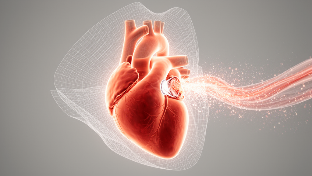
Mitral regurgitation, the backward leak of blood through the heart’s mitral valve, is the most common valvular heart disease in the United States and the second most common in Europe, affecting an estimated 3 to 6 percent of the global population. Even when the left ventricle is pumping normally, moderate and severe regurgitation carries a measurable excess mortality risk, which makes accurate and reproducible assessment of the leak a matter of life-and-death timing for diagnosis and treatment. A new review published in the journal Cardiovascular Ultrasound examines how the field is moving from decades-old geometric approximations toward artificial intelligence tools that measure regurgitant flow directly, frame by frame, across the whole of systole.
Echocardiography remains the foundational investigation for mitral regurgitation because of its high accessibility, but its standard quantification methods rest on assumptions that real hearts rarely obey. Among the traditional qualitative, semi-quantitative and quantitative measures, the effective regurgitant orifice area and the regurgitant volume are the most predictive of clinical outcomes, and both are typically derived using the proximal isovelocity surface area method, known as PISA. That method, despite its widespread use in clinical practice and in landmark trials, suffers from three well-documented shortcomings: it underestimates severity when there are multiple or eccentric jets, it relies on a single snapshot within the cardiac cycle, and it assumes the regurgitant orifice is circular. These limitations matter because a single central jet with a circular orifice is rarely present in reality. Other conventional parameters, such as color flow of the regurgitant jet, the continuous wave regurgitant jet profile and pulmonary vein flow, are simple and easily acquired, but they too are susceptible to inaccuracy when the regurgitant jet is eccentric.
Recent advances in artificial intelligence and deep learning have produced automated and semi-automated tools designed specifically to address the limitations of the assumed single, spherical, holosystolic jet. One line of work targets the PISA measurement itself. Automated measurements of PISA using deep learning allow real-time, multi-frame assessment, something previously impractical in clinical practice because manual measurement in every frame was prohibitively laborious. A deep learning framework called EasyPISA, published in 2024, demonstrated that multi-frame automatic segmentation of the flow convergence region from two-dimensional color Doppler was possible without hemispherical assumptions, showing strong agreement with manual transthoracic echocardiographic and cardiac magnetic resonance assessment of MR severity. The review’s authors are careful to note the caveats: this proof-of-concept application was validated in a retrospective cohort, is not commercially or widely available, and prospective testing is planned.
A parallel approach extends PISA into three dimensions. Three-dimensional PISA offers improved image quality and greater accuracy, particularly in regurgitation that is eccentric, significant, or arises from an asymmetrical orifice, precisely the situations in which the geometric assumptions of the two-dimensional method break down. Automated three-dimensional PISA detection has been shown to require minimal training in 3D software analysis, to reduce the time spent performing measurements, and to achieve good reproducibility, potentially reducing inter-operator variability even among less experienced operators. Its short postprocessing time, limited to a few minutes, makes it relatively practical for routine clinical use. However, the initially available commercial implementation, Siemens’ eSie PISA, is no longer widely available.
The most technically detailed newcomer is 3D Auto Color Flow Quantification, a Philips algorithm validated against cardiac magnetic resonance imaging. Unlike conventional methods, it does not use a single baseline-shifted color velocity and does not assume a single circular orifice producing a perfectly hemispherical isovelocity shell. In fact, the algorithm makes no geometric assumptions at all about the size, shape or constancy of the orifice. Its fluid model is grounded in the Navier-Stokes equations describing the motion of fluid in space and time, with the dynamic color 3D jet segmented from a time sequence of stabilized 3D ultrasound images. Mathematical morphology is then used to estimate the average orientation of the jet over the systolic frames, and that orientation guides the search for the regurgitant orifice in each image independently.
From there, the algorithm builds a dynamic surface model of the valve. It locates the mitral valve along the previously determined jet directions and applies an analytic geometric regression to fit a surface model, from which a dynamic regurgitant orifice is identified across the entire time sequence of stabilized 3D ultrasound images. The four-dimensional ultrasound Doppler color data are then searched through the color volume along that valve surface to identify one or more regurgitant orifices. Finally, a flow convergence model is dynamically fit to the image sequence based on the location of the identified orifice or orifices and the surrounding color data. The overall model is a convolution of many smaller pinhole flow models whose individual flow contributions are summed to equal the total flow in the color dataset. Because several orifices, or a complex orifice of various shapes such as a slit, are handled natively, the method is not tied to the pinhole, circular-orifice assumption embedded in the current standard-of-care PISA method. Validation work has included data from primary and secondary mitral regurgitation, single and multiple jets, and both concentric and eccentric jets.
Obtaining a 3D Auto CFQ measurement requires four distinct steps: image acquisition, mitral valve model creation, review of the flow model, and flow analysis to obtain the regurgitant volume. Acquisition begins with a high-quality 3D color Doppler dataset, and because the software analyzes dynamic temporal changes in flow throughout systole, a volume rate of at least 20 hertz is essential for sufficient temporal resolution. When higher frame rates are difficult to achieve, as in patients with arrhythmia or rapid heart rates, multi-beat acquisition can be used. A high color Doppler scale is also critical, with a minimum Nyquist limit of roughly 70 to 75 centimeters per second recommended, achievable by reducing the color frequency to 3.3 megahertz. Suboptimal velocity settings, particularly low color scales, can cause signal saturation, aliasing and underestimation of regurgitant flow. To support quality assurance, the software provides a data adequacy indicator that alerts the operator to suboptimal volume rate or aliasing velocity settings in real time.
The remaining workflow steps blend automation with operator oversight. Within the software tool, a dynamic 3D greyscale model of the mitral valve is created, providing the framework for identifying the regurgitant orifice. Automated landmark detection covers the annulus, the anterior and posterior leaflets and the coaptation zone, but the operator can adjust these if required; the static model defines valve structure at a single reference point while the dynamic model captures valve motion throughout systole. The software then displays the regurgitant flow within the 3D color Doppler volume and enables exclusion of confounding signals such as aortic outflow. If misalignment, inaccuracies or artefact are detected, the operator can return to previous steps to refine annular landmarks, leaflet tracking or dynamic motion before proceeding. Once the model is confirmed, the software performs automated quantification, analyzing each 3D color Doppler frame as individual pinholes of regurgitation that are summed into the total regurgitant volume in milliliters, so that the size, shape and number of orifices are accounted for rather than assumed.
Important limitations remain. The current 3D MR flow quantification software has been shown to yield a smaller regurgitant volume compared with two-dimensional PISA, which means the optimal cutoff value for severe MR may differ from thresholds established with other methods, and the review stresses that further studies are needed for better validation. Separately, other deep learning frameworks have been developed to identify the presence and severity of MR directly from four-chamber and/or two-chamber transthoracic echocardiographic clips, generally relying solely on qualitative measures based on color Doppler videos. Rather than delivering a more accurate diagnosis, the authors suggest these tools may be most useful for screening, especially in resource-poor settings where cardiologist review of every echocardiographic image is not feasible, flagging patients who would benefit from further evaluation using established quantitative methods such as measuring effective regurgitant orifice area and regurgitant volume.
The review concludes that artificial intelligence-based quantification of mitral regurgitation is time-efficient and addresses the false geometric and temporal assumptions intrinsic to traditional techniques. Because AI models have shown low inter-observer and intra-observer variability, incorporating automated measurements could assist clinicians as a second check in challenging cases, help monitor disease stability or progression over time, and streamline workflows by reducing reporting time. Three-dimensional Auto CFQ in particular enables automated frame-by-frame assessment that may improve accuracy and reproducibility and reduce operator dependency while maintaining workflow efficiency. Early data show strong concordance with cardiac magnetic resonance, yet current algorithms tend to yield smaller regurgitant volumes than conventional PISA-based approaches. The authors’ bottom line is measured: while integrating AI-based quantification into routine echocardiographic practice may improve diagnostic accuracy and enable improved longitudinal monitoring in patients with MR, further studies are required to validate these measurements before they can reshape everyday care.
Subject of Research: Artificial intelligence methods for quantifying mitral regurgitation on echocardiography
Article Title: Quantification of mitral regurgitation: from traditional methods to artificial intelligence
Article References: Cho, K., Su, J., Bonnefous, O., Prabhu, S., & Anthony, C. (2026). Quantification of mitral regurgitation: from traditional methods to artificial intelligence. Cardiovascular Ultrasound, 24(1), Article 14. https://doi.org/10.1186/s12947-026-00374-6
Image Credits: AI Generated
DOI: 10.1186/s12947-026-00374-6
Keywords: mitral regurgitation, echocardiography, artificial intelligence, deep learning, 3D Auto CFQ, PISA, cardiac MRI, regurgitant volume, valvular heart disease, color Doppler, medical imaging, machine learning
News Source: Ophelia Keating. (October 29, 2026). AI Takes On the Flawed Math of Measuring Leaky Heart Valves. Scienmag.



