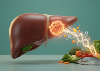
A team of researchers at Gannan Medical University in China has combined bulk RNA sequencing, single-nucleus sequencing and machine learning to map the molecular and cellular landscape of liver fibrosis in metabolic dysfunction-associated steatohepatitis, or MASH. Their integrative analysis, published in Frontiers in Cell and Developmental Biology, produced a compact seven-gene signature that distinguishes advanced from mild fibrosis in an independent patient cohort, and identified specific liver cell subpopulations and signaling axes that appear to accompany the progression of scarring in this increasingly common liver disease.
MASH, previously known as non-alcoholic steatohepatitis, is the severe inflammatory form of metabolic dysfunction-associated steatotic liver disease. It is characterized by fat accumulation, inflammation and, critically, liver fibrosis driven by collagen-producing hepatic stellate cells. Fibrosis stage is the major predictor of mortality in these patients and the main determinant of progression toward cirrhosis and hepatocellular carcinoma, yet effective therapies to reverse MASH-associated fibrosis remain lacking. Because the molecular mechanisms that push fibrosis forward are incompletely understood, the researchers set out to build a comprehensive, multi-layered picture of the transcriptomic changes involved.
The discovery work began with four publicly available human bulk RNA-seq datasets covering 254 MASH patients with mild fibrosis, classified as stage F0 to F2, and 122 patients with advanced fibrosis, stage F3 to F4. After removing low-expression genes and outliers, the team used the ComBat method to correct batch effects arising from different sequencing protocols and merged the data into a single cohort of 331 individuals: 219 with mild fibrosis and 112 with advanced fibrosis. Differential expression analysis on this merged cohort identified 488 genes upregulated and 125 genes downregulated in advanced fibrosis, with the upregulated genes strongly enriched for extracellular matrix organization, collagen fibril organization and related structural processes, while downregulated genes were linked to lipid and amino acid metabolism.
To connect these gene-level changes to coordinated biological programs, the researchers applied weighted gene coexpression network analysis to the 5,000 most variable genes across the merged cohort. This yielded ten gene modules, four of which correlated significantly with both MASH score and fibrosis grade. Two stood out: a 347-gene module centered on extracellular matrix organization and a 74-gene module tied to epithelial cell morphogenesis. Gene set enrichment analysis confirmed that genes from both modules were most highly enriched among those upregulated in advanced fibrosis, marking them as the network-level fingerprints of progressing disease.
The team then narrowed the field. Robust rank aggregation across datasets, combined with the merged-cohort differential expression results and the key network modules, produced an overlapping set of 74 candidate genes. Three machine learning algorithms—LASSO regression, random forest and support vector machine-recursive feature elimination—were applied in parallel, and the intersection of their selections left seven hub genes: CAPN6, FAP, KRT23, SOX4, STMN2, TAGLN and VWF. In an independent external cohort of 56 MASH patients, this seven-gene panel achieved an area under the receiver operating characteristic curve of 0.81, with a 95 percent confidence interval of 0.69 to 0.93, for discriminating advanced from mild fibrosis. That performance was close to the 74-gene panel’s AUC of 0.84 in the same cohort, showing that the streamlined signature largely preserves discriminative power. Expression of the seven genes also rose with fibrosis stage in the separate SteatoSITE database of 940 histologically confirmed patients.
Not every gene behaved identically across settings, a nuance the authors report candidly. When sex was included as a covariate in the discovery cohorts, KRT23 and STMN2 lost significance in one dataset and STMN2 and TAGLN in another, an attenuation the researchers attribute to the small proportion of male patients in the advanced fibrosis groups of those cohorts. In a cross-species check, mice fed a methionine-choline-deficient diet for six weeks showed significantly increased liver mRNA expression of Capn6, Fap and Krt23, but Sox4, Stmn2, Tagln and Vwf showed no significant transcript-level differences in this model, and two borderline results disappeared after Bonferroni correction. The authors also note that the MCD diet captures diet-driven steatohepatitis rather than the full metabolic context of human MASH, so the roles of these genes warrant further investigation.
To find out which cells generate the fibrosis-associated signals hidden in bulk tissue, the researchers turned to a single-nucleus RNA-seq dataset of liver biopsies from 31 MASH patients across fibrosis stages, from which they recovered roughly 50,000 high-quality nuclei representing hepatocytes, cholangiocytes, endothelial cells, macrophages, hepatic stellate cells and several immune populations. Deconvolution of the bulk merged cohort using this nuclear reference showed that the extracellular matrix and epithelial morphogenesis modules correlated positively with hepatic stellate cells and cholangiocytes, while the immune response module tracked macrophages and the protein-targeting module tracked hepatocytes. Within the stellate cell compartment, one subcluster expressing the activation markers COL1A1 and TIMP2 emerged as the highly fibrogenic activated stellate population, its upregulated genes enriched for matrix organization and collagen fiber assembly.
Two further subpopulations drew particular attention. Among cholangiocytes, one subset marked by FGF13 and PPARGC1A expression showed the strongest interactions with hepatic stellate cells of any cholangiocyte cluster, exchanging signals including VEGFA with its receptor NRP1 and APP with CD74. Transcription factor regulon analysis identified CREB5 as a key regulator specifically active in this FGF13-positive subset, with target genes enriched in immune signaling pathways and epithelial-mesenchymal transition; CREB5 expression was elevated across multiple MASH animal models and showed a generally increasing trend in human MASH. Among hepatocytes, a subset defined by high expression of the acute-phase genes SAA1 and SAA2 engaged in prominent communication with stellate cells through the RSPO3-LGR4 and IGF1-IGF1R axes and with macrophages through TNFSF14-TNFRSF14 and SELE-CD44. STAT3 emerged as the leading regulator of this SAA-positive hepatocyte population, its target genes enriched in complement activation and inflammatory pathways, and its regulatory activity correlating most strongly with SAA1 and SAA2 expression.
The authors are explicit that these are associative, bioinformatic findings rather than demonstrated mechanisms. They acknowledge several limitations: residual batch effects may persist despite correction, the analysis relies solely on transcriptomics without multi-omic integration, using a single-nucleus reference to deconvolve whole-tissue bulk data introduces a nuclear-versus-cytoplasmic transcriptome mismatch, and single-nucleus data lack spatial context. Most importantly, no functional experiments in cells or living systems validate the proposed regulatory roles, which the researchers identify as the primary limitation of the work; the mouse validation also used only male animals.
Even with those caveats, the study offers a concrete resource for a field where fibrosis stage drives clinical outcomes. The seven-gene tissue signature could support patient stratification and cross-cohort reproducibility in translational research, while the mapped cell subpopulations, transcription factors and ligand-receptor pairs—CREB5 in FGF13-positive cholangiocytes, STAT3-driven SAA production in hepatocytes, and the RSPO3-LGR4, IGF1-IGF1R and VEGFA-NRP1 axes linking hepatocytes, cholangiocytes, stellate cells and macrophages—provide a ranked set of hypotheses for the functional experiments that must follow before any of these molecules can be considered therapeutic targets.
Subject of Research: Integrative transcriptomic analysis of biomarkers and cell types associated with liver fibrosis progression in MASH
Article Title: Integrative machine learning analysis of bulk and single-nucleus RNA sequencing reveals key cell types and biomarkers driving MASH-associated fibrosis
Article References: Zhang, J., Wang, J., Shen, Y., Zhang, Y., Luo, Z., Kong, X., Li, W., Luo, J., Xu, F., & Wan, S. (2026). Integrative machine learning analysis of bulk and single-nucleus RNA sequencing reveals key cell types and biomarkers driving MASH-associated fibrosis. Frontiers in Cell and Developmental Biology, 14, Article 1961567. https://doi.org/10.3389/fcell.2026.1961567
Image Credits: AI Generated
DOI: 10.3389/fcell.2026.1961567
Keywords: MASH, liver fibrosis, RNA sequencing, machine learning, biomarkers, hepatic stellate cells, cholangiocytes, hepatocytes, single-nucleus sequencing, gene signature, cell-cell communication, transcription factors
News Source: Juliet Wilcox. (October 29, 2026). Machine Learning Pinpoints Seven Genes and Key Liver Cells Behind MASH Fibrosis. Scienmag.