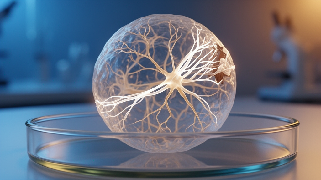
For decades, one of the most stubborn problems in neurology has been the absence of any approved therapy that helps the brain rebuild its myelin, the fatty insulation that wraps around nerve fibers and allows electrical signals to travel at speed. When myelin is destroyed, as happens in multiple sclerosis, the consequences can be devastating: slowed signaling, progressive disability and, ultimately, irreversible neurodegeneration. Now, a team of researchers working across Australian and New Zealand institutions has unveiled a laboratory model that, for the first time, allows scientists to watch human myelin being damaged and then repaired in a dish, cell by cell and molecule by molecule. The work, published in Nature Neuroscience, describes spheroids grown from human induced pluripotent stem cells that contain mature, myelinating oligodendrocytes alongside functionally reactive microglia, the immune cells of the central nervous system.
The technical achievement behind the model is considerable. The researchers began with established protocols for generating spinal cord-patterned spheroids, which require seven weeks of neural patterning followed by roughly twelve weeks of maturation, during which time oligodendrocytes extend their myelin sheaths around axons. Crucially, they then added a second ingredient: macrophage-like precursor cells, themselves derived separately from induced pluripotent stem cells, which were seeded into the spheroids three weeks into the maturation phase. This step was deliberately designed to mimic human development, in which microglial progenitors migrate into the central nervous system after neurogenesis has begun. Over subsequent weeks, through intrinsic signaling within the spheroids, these macrophage-like cells differentiated into bona fide microglia, losing CD45 expression and enriching the microglial markers P2RY12 and CX3CR1. Live imaging of fluorescently labeled cells showed the newcomers integrating into the tissue and extending and retracting their ramified processes, dynamically surveying their environment just as microglia do in a living brain.
With the cellular cast assembled, the team set about inflicting a controlled injury. They applied lysolecithin, a detergent-like compound that disrupts lipid membranes and is a mainstay of demyelination studies in rodents, at a concentration of 0.1 milligrams per milliliter for sixteen hours. The effect was unmistakable. Myelin basic protein staining revealed extensive fragmentation of myelin sheaths, and the colocalization between myelin and neurofilament-heavy-labeled axons dropped sharply. An automated analysis pipeline, which measured the density of myelin sheaths longer than twenty micrometers per unit area of axon, confirmed a significant demyelinating effect of the treatment. Transmission electron microscopy provided ultrastructural confirmation, showing axons wrapped in visibly damaged myelin. The team also observed disruption of paranodal assembly, with the oligodendrocyte protein claudin-11 dispersing along demyelinated axons, a phenomenon that echoes findings in postmortem tissue from multiple sclerosis patients.
What happened next is where the model truly earns its keep. Within hours of the injury, the microglia responded. Single-cell analysis of multiplexed immunolabeling revealed a significant increase in HLA-DR, a marker of antigen presentation and microglial activation, at twenty-four hours and seven days after the insult, with effect sizes exceeding 0.8. More strikingly, by seven and fourteen days after lysolecithin removal, the researchers documented significant colocalization of myelin debris within IBA1-positive microglia, suggesting active phagocytosis. To be certain, the team turned to correlative light and electron microscopy, a technique that allows the same individual cell to be tracked from fluorescence imaging down to nanometer-scale ultrastructure. The electron micrographs showed microglial cytoplasm packed with lysosomal structures filled with electron-dense material consistent with ingested myelin, along with the vacuolization and membrane whorls characteristic of phagocytic cells. In parallel, the microglia engulfed latex beads in two-dimensional culture and secreted interleukin-6, tumor necrosis factor and interleukin-10 when stimulated with lipopolysaccharide, further validating their immune functionality.
To connect the cellular observations to human disease, the researchers performed single-cell RNA sequencing on spheroids twenty-four hours after the demyelinating insult. The dataset, spanning more than ninety thousand cells after quality control, revealed that oligodendrocytes in the injured spheroids upregulated genes involved in ion channel regulation and cellular stress, including KCNMB4 and HSPB1, both of which are known to be upregulated in human multiple sclerosis lesions. Oligodendrocyte precursor cells showed a dramatic transcriptional shift, with 637 genes upregulated and 2,708 downregulated, including activation of cell division pathways and immune-related genes such as IRF9 and CDKN1A. Microglia shifted toward lipid metabolism and antigen presentation pathways. When the team correlated their spheroid data with published gene expression profiles from active multiple sclerosis lesions in human postmortem tissue, the association was statistically significant, and the overlap between upregulated genes in the spheroid oligodendrocytes and those in two independent human lesion datasets was highly significant by Fisher’s exact test. In other words, the dish was not merely resembling disease superficially; it was recapitulating molecular signatures of human pathology.
The central question, however, was whether the spheroids could repair themselves. To answer it, the researchers supplemented the culture medium with ethynyl deoxyuridine, a thymidine analog that is incorporated into the DNA of dividing cells. Any oligodendrocyte carrying this label must have been generated after the injury, since surviving mature oligodendrocytes are postmitotic. The results were definitive. The proportion of newly generated oligodendrocytes rose significantly at four, eight and twelve weeks after lysolecithin removal, and by eight weeks a significant fraction of these newborn cells had commenced forming myelin sheaths. The density of myelin sheaths along axons returned to levels indistinguishable from uninjured controls, and paranodal structures marked by claudin-11 reappeared, indicating mature, compact myelin. Focused ion beam scanning electron microscopy, a technique capable of resolving individual membrane layers at nanometer resolution, confirmed the presence of compact myelin wrapped around axons in the repaired spheroids.
The ultrastructural quantification also revealed a well-known signature of repair: the G-ratio, which measures the relationship between axon diameter and total fiber diameter, was significantly higher in the repaired spheroids than in uninjured controls. A higher G-ratio means thinner myelin, and thinner myelin is precisely what is expected during early remyelination, both in animal models and in human tissue. This finding carries particular scientific weight because it addresses a long-standing debate in the field. Evidence from carbon-14 isotope birthdating of oligodendrocytes in human shadow plaques had suggested that human remyelination might occur only through oligodendrocytes that survive the demyelinating insult. The new data demonstrate directly that human oligodendrocyte precursor cells, at least in this model system, retain the capacity to proliferate, differentiate and generate new myelinating oligodendrocytes after injury, providing in vitro evidence for a mechanism that could not previously be observed in human tissue.
The model also passed a practical test that matters enormously for drug development. When the researchers treated injured spheroids with clemastine fumarate, an antihistamine that was the first putative remyelinating drug to enter clinical trial for multiple sclerosis, they observed a significant increase in the proportion of newly generated oligodendrocytes that went on to myelinate, compared with injured spheroids cultured in base medium alone. No such effect appeared in vehicle-treated spheroids. This result establishes the platform as a viable drug-screening tool, one that can validate candidate therapies in a human cellular context before they are advanced toward expensive and often disappointing clinical trials. The authors acknowledge that the system, given its cellular complexity and long culture times, is not suited to high-throughput screening in its current form, but its physiological relevance makes it ideal for validating a refined set of drug candidates or for genetic manipulation.
The researchers are candid about the model’s limitations. Induced pluripotent stem cell-derived organoids are known to mirror a fetal rather than adult brain in transcriptional maturity, a caveat given that multiple sclerosis largely affects adults. Organoid-to-organoid variability is substantial, and only about half of the differentiations successfully remyelinated, with those failures tending to correspond to differentiations that had not myelinated well in the first place. Of twenty-nine independent differentiations of the primary cell line, sixty-nine percent produced myelin, ranging from robust to moderate. The team compensated by combining technical and biological replicates across numerous spheroids and differentiations, and they suggest that fluorescence reporter lines could eventually allow enrichment of highly myelinated spheroids before assays begin. Even with these caveats, the platform offers something no other human system can: the ability to quantify compact myelin ultrastructure in the context of active repair, to observe microglia clearing debris in real time, and to trace the birth of new myelinating cells after injury. For a field that has watched countless remyelination therapies succeed in mice and fail in humans, that combination of human relevance and mechanistic depth may prove to be exactly the tool needed to finally translate myelin repair from promise into medicine.
Subject of Research: A human induced pluripotent stem cell-derived spheroid model for studying central nervous system myelin injury and remyelination relevant to multiple sclerosis
Article Title: A human-induced pluripotent stem cell-derived spheroid model to investigate myelin injury and repair
Article References: Ramesan, S., Mason, J., Kumar, S. S., Swanson, M. E. V., Korneev, D., Mills, S. A., Mathangasinghe, Y., Andres, A., Loh, L., Fraser, T. D., Lewis, K. N., Beau, F.-X. E., Chua, S., Kapoor-Kaushik, N., Williams, L. J., Mrkela, M., Turner, B. J., Ramm, G., Rutar, M. V., … Barton, S. K. (2026). A human-induced pluripotent stem cell-derived spheroid model to investigate myelin injury and repair. Nature Neuroscience. https://doi.org/10.1038/s41593-026-02457-z
Image Credits: AI Generated
DOI: 10.1038/s41593-026-02457-z
Keywords: multiple sclerosis, myelin, remyelination, oligodendrocytes, microglia, induced pluripotent stem cells, spheroids, demyelination, organoids, drug screening, neuroinflammation, single-cell RNA sequencing
News Source: Cassandra Pierce. (October 9, 2026). Lab-grown human brain spheroids show myelin can repair itself after injury. Scienmag.