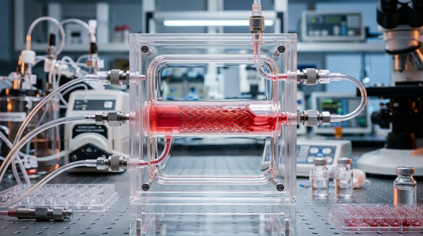
Engineers have built a silicone blood vessel lined with living human endothelial cells that is large enough to hold a real stent, and they have shown that the model can reproduce key cellular responses to blood flow while quantifying how cells recolonize the metal mesh after implantation. The work, published in BMC Methods, provides detailed fabrication protocols for a cell-cultured polydimethylsiloxane (PDMS) vascular model designed to allow placement of implant devices such as self-expanding stents under controlled flow conditions.
The motivation comes from a persistent problem in interventional cardiology. A stent maintains blood flow by expanding a stenotic artery from the inside, but current stents are mechanically sub-optimized and can exert excessive forces on the vascular wall. That excessive mechanical stimulation can trigger inflammation, late thrombosis, and in-stent restenosis, and the mismatch between stent stiffness and vessel stiffness remains unresolved. Optimizing stent mechanics requires reproducing the mechanical field within the stented vessel and evaluating endothelialization, the process by which endothelial cells adhere, migrate, and form a confluent monolayer on the stent surface, which serves as a key biological indicator of vascular neointimal formation.
Existing tools fall short in different ways. Numerical simulations using patient-specific or animal-derived data have provided valuable insights into stress and strain distributions in stented vessels, but they cannot capture the cellular-scale events that determine vascular compatibility. Studies that culture endothelial cells on surfaces containing stent struts can assess surface biocompatibility, but they lack the realistic mechanical environment to which stents and blood vessels are exposed. Perfusion systems with small-to-medium vascular models have enabled detailed analysis of endothelial cell dynamics in stented vessels, yet their fabrication processes remain complex and technically demanding, which limits reproducibility and accessibility for broader application.
The new model addresses this gap with a straightforward construction route. The team fabricated a straight channel vascular model with a circular cross-section from PDMS, a silicone elastomer used to mimic the adventitial structure of arteries. Connectors serving as the inlet and outlet were cast in polyacetal molds, bonded, and cured at 60 degrees Celsius. A stainless-steel core rod was then inserted through the connectors, PDMS was poured into the surrounding mold, degassed, and cured, and the core rod was finally removed by immersing the cured model in anhydrous ethanol, whose low surface tension and high volatility allow it to penetrate the interface and release the rod with minimal force. Three model sizes were produced, with inner and outer diameters of 4 and 5 millimeters, 5 and 6 millimeters, and 6 and 7 millimeters.
Dimensional checks showed the fabrication method is reasonably precise. Each model was cut at five locations and measured under a stereomicroscope. The deviation from the target inner diameter was within plus or minus 100 micrometers for each model, and wall thickness deviations were within plus or minus 150 micrometers for the 4-millimeter models, plus or minus 250 micrometers for the 5-millimeter models, and plus or minus 200 micrometers for the 6-millimeter models. Inner diameter precision depends strongly on the accuracy of the core rod, while wall thickness variation increases with larger diameters because the connector bonding area shrinks and misalignment during fabrication becomes more likely. The developed models have a wall thickness of 500 micrometers, which is comparable to the average thickness of medium-sized blood vessels such as carotid and coronary arteries.
To create the living lining, the researchers seeded human carotid artery endothelial cells (HCtAECs) onto the lumen of the 4-millimeter model. The interior was first hydrophilized with plasma treatment and coated with a gelatin and poly-D-lysine mixture to promote cell adhesion. A cell suspension at a concentration of 5.0 times 10 to the 5 cells per milliliter was injected, and the model was held at a series of tilt angles during incubation so the cells could settle evenly around the circumference. After 24 hours, the cells formed a confluent monolayer covering the lumen, with the adhesion protein VE-cadherin visible at intercellular junctions and no gaps in the endothelium. In static culture, however, most cells detached by 48 hours after seeding, because growth factors in the medium allowed endothelial proliferation to continue past confluence, disrupting the monolayer.
Flow proved essential for maintaining the lining. The cultured model was connected to a flow circuit with a peristaltic pump and two reservoirs, kept at 37 degrees Celsius, and the cells were exposed to two levels of fluid shear stress for up to 48 hours: 0.5 pascals at a flow rate of 4.19 milliliters per second, corresponding to laminar flow, and 2.3 pascals at 8.38 milliliters per second, where the Reynolds number exceeded 2,300 and the friction coefficient was estimated with the Blasius equation. Under shear stress, the cells elongated and oriented in the direction of flow, developed actin stress fibers parallel to the flow, and showed patchy VE-cadherin distribution at adhesion sites, consistent with migration and shape change. Quantitative analysis of aspect ratio and orientation angle, based on at least three independent experiments with 50 cells extracted per experiment, confirmed significant elongation and flow alignment. The researchers also found no difference in the cellular response between the upper and lower sides of the large-diameter model, indicating that gravity’s influence could be ignored.
Shear stress magnitude affected cell retention. The detachment rate, defined as the area of cell loss relative to the total luminal surface area in the images, increased significantly under the 0.5-pascal condition, averaging just under 20 percent, while the static and 2.3-pascal conditions remained at approximately 10 percent. The authors note that 0.5 pascals represents sub-mean shear stress in arteries and is close to values observed at atherosclerotic lesion sites downstream of endothelial dysfunction, whereas the 2.3-pascal condition falls within the roughly 1 to 2 pascal range reported for human carotid artery walls. Low shear stress has been reported as a risk factor for endothelial dysfunction, which the authors suggest could be induced by similar mechanisms in their model. The cultured vascular model maintained its performance for at least 96 hours after cell seeding.
The central demonstration was stent placement during perfusion culture. A self-expanding stent made of nickel-titanium shape memory alloy, with an outer diameter of 6 millimeters, was shrunk to approximately 4 millimeters by cooling in ice water, mounted in an industrial straw, sterilized in 70 percent ethanol, and inserted into the cultured model through a T-shaped fitting after temporarily stopping the flow. Shear stress of 2.3 pascals was then reapplied, and the cells were cultured for up to 48 hours. Endothelial cells migrated from the lateral to the top sides of the stent between 24 and 48 hours after placement. On the lateral side, the average endothelialization rate was just over 20 percent at 24 hours and increased to approximately 50 percent at 48 hours, while the rate on the inner top side stayed below 10 percent, increasing only slightly. Complete coverage of the inner top side, the authors conclude, requires further long-term perfusion culture.
The authors present the platform as a foundation for stent design rather than a finished replica of an artery. The model reproduces the mechanical field of a stented vessel, including the radial and circumferential forces generated during stent expansion, which stent struts simply placed on a culture surface cannot, and its optical transparency allows observation of cells compressed between struts and the wall. But the limitations are explicit: the biological tissue exhibiting vascular function is only an endothelial monolayer, without the smooth muscle cell crosstalk needed to reproduce neointimal hyperplasia; the 500-micrometer PDMS wall is far stiffer than a real vessel, so the stent did not sink into the wall and vessel diameter changes were minimal, meaning the mechanical field was not fully reproduced; and the forces between stent and wall have not yet been quantitatively measured. The team proposes thinner walls, softer materials such as alginate hydrogel, stress visualization techniques, and complex geometries with curvatures and bifurcations as future directions, with the aim of quantifying the relationship between stent-induced mechanical forces and restenosis to determine optimal stent stiffness.
Subject of Research: An in vitro PDMS vascular model lined with human endothelial cells for evaluating stent placement and endothelialization under controlled shear stress
Article Title: Cell-cultured PDMS vascular model to allow placement of implant devices
Article References: Okuno, T., Ito, K., Funamoto, K., & Yoshino, D. (2026). Cell-cultured PDMS vascular model to allow placement of implant devices. BMC Methods, 3(1), Article 16. https://doi.org/10.1186/s44330-026-00072-9
Image Credits: AI Generated
DOI: 10.1186/s44330-026-00072-9
Keywords: stent, endothelialization, PDMS vascular model, shear stress, endothelial cells, in-stent restenosis, perfusion culture, tissue engineering, biomedical engineering, vascular stenosis, NiTi stent, flow circuit
News Source: Gregory Coleman. (November 4, 2026). Lab-grown blood vessel model lets researchers test stents under real flow. Scienmag.