The fibrocartilaginous disc that cushions the jaw joint in every mammal that has one appears to have begun its evolutionary life as a tendon.
The mammalian jaw joint is an evolutionary innovation. Where non-mammalian jawed vertebrates articulate the lower jaw through the primary joint between the articular and quadrate bones, mammals hinge the dentary bone of the lower jaw directly against the squamosal, part of the temporal bone of the skull. This dentary-squamosal joint, documented in the fossil record as mammaliaforms expanded the dentary to make contact with the squamosal, is thought initially to have acted as a buttress stabilising the older primary joint and preventing dislocation, before taking over as the dominant joint. It comprises the mandibular condyle, the glenoid fossa it articulates against, and, interposed between them, the articular disc.
That disc matters far beyond evolutionary curiosity. It divides the joint into superior and inferior cavities, cushions the articulating surfaces, and its pathology underlies several temporomandibular disorders. Recent meta-analyses and systematic reviews report that TMD affects roughly 31 percent of adults and elderly patients under standard diagnostic criteria. Yet despite extensive knowledge of disc morphology and biomechanics, the genetic, cellular and molecular mechanisms of disc development have remained poorly understood, a gap that also hampers disc regeneration strategies.
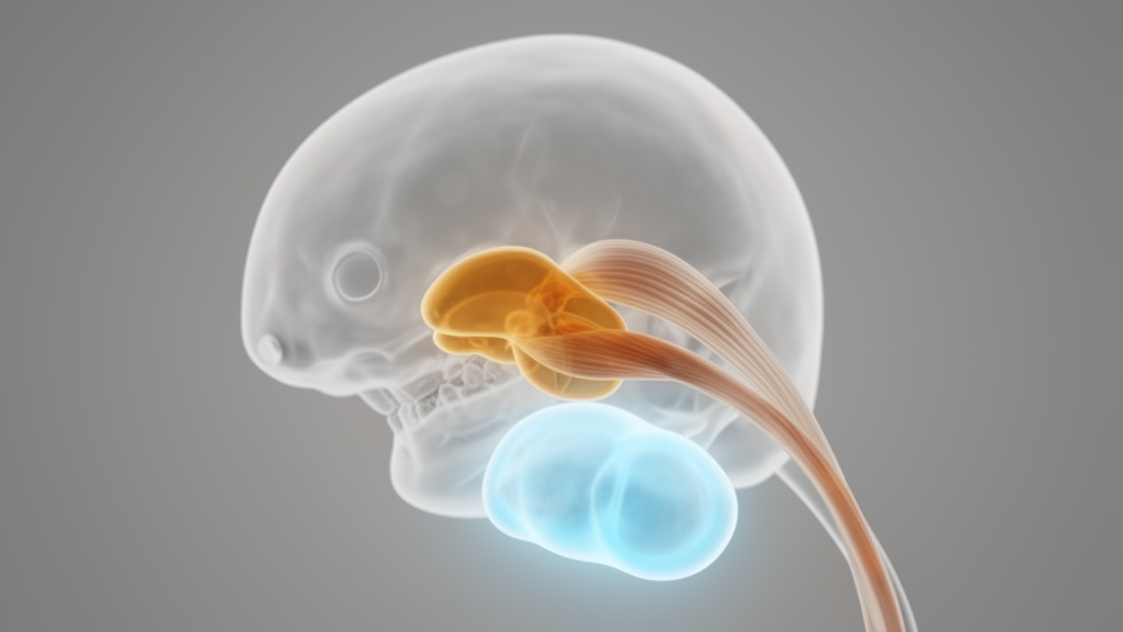
Two competing hypotheses dominated. One held that the disc originates from the superficial layer of the condylar cartilage, a plausible idea given that both structures are fibrocartilaginous and lie in intimate contact during development. The other proposed that the disc forms independently, from its own mesenchymal condensation, and a further suggestion, based on the insertion of the lateral pterygoid muscle and the discomalleolar ligament into the disc, was that the disc began as a tendon associated with that muscle, which became trapped between the newly abutting dentary and squamosal bones as the mammalian jaw joint evolved.
To resolve the question, Ticha Tuwatnawanit, Ying Sun, Neal Anthwal and Abigail Tucker of King’s College London analysed the condyle and disc throughout murine development, combining bulk RNA sequencing of laser-captured tissue, clonal lineage tracing and mouse knockouts. The condyle emerges as a secondary cartilage associated with the dentary at embryonic day 14.5. At that stage the disc was not yet morphologically distinct, but the team found that the gene Lgr5, a marker of forming joint interzones, was already expressed in a band above the Sox9-positive condylar primordium. Strikingly, even at E13.5, before the condyle is induced, a distinct band of Lgr5 was evident in the region of the future joint, and this domain overlapped with expression of the tendon marker Scleraxis, highlighting the tendon-like character of these earliest disc cells.
By E16.5 a nascent disc was visible on histology atop the condyle as the upper joint cavity formed, and Lgr5 expression remained prominent in the forming disc. By E18.5 the disc had lifted off the condyle with formation of the lower cavity, and Lgr5 was still strongly expressed in disc cells. Sox9 expression, by contrast, had switched off in the superficial condylar layers while remaining robust in the chondrogenic layers beneath. Using these two markers, the disc and condyle could be distinguished as separate primordia from the onset of joint development.
The transcriptomic analysis reinforced the distinction. Laser capture microdissection isolated disc and condyle tissue from Scleraxis-Cre;TdTomato embryos at E14.5, E15.5 and E16.5 for bulk RNA sequencing. Principal component analysis showed that samples clustered strongly by developmental stage, while the disc profile nested largely within the condyle profile, reflecting the shared fibrocartilaginous identity of both structures. But differential expression analysis told a sharper story: as development proceeded, osteogenic and chondrogenic genes were upregulated in the forming condyle, while the disc became enriched for tenogenic genes including Postn, Tnn, Ptn, Thbs2, Mfap4 and Bgn, all at low levels in the condyle. At E16.5 the disc expressed collagen type I genes but little of the osteoblast markers Alpl, Ibsp, Spp1 and Bglap that rose in the condyle. The disc also increased Sox9, Aggrecan and Wwp2, consistent with its fibrocartilage designation, but its trajectory was unmistakably tendon-like.
Clonal tracing provided perhaps the most decisive evidence. Using Pcag-CreER;Confetti mice, in which a single tamoxifen dose at E14.5 stochastically labels cells with one of four fluorescent colours, the researchers labelled cells before the disc and condyle physically separated, then examined the joint at E18.5. Neighbouring cells sharing a colour were counted as clones. Disc and condyle each contained their own distinct clones, and no clone spanned the boundary between the disc and the superficial condylar layer. Condylar surface clones divided along the surface and into the cartilage, but disc and condyle cells simply did not mix. After their establishment, the two populations behave as separate lineages, arguing strongly that disc cells are not drawn from condylar cartilage.
A final genetic test asked whether the disc can even begin without its neighbour. In Wnt1-Cre;Sox9fl/fl mutants, where Sox9 is deleted in neural crest-derived tissue, the condyle fails to form entirely. Nevertheless, at E15.5 and E18.5 the mutants retained an Lgr5-positive band of cells above the membranous dentary bone, in the position of the disc, even though the condyle and much of the fossa were disrupted. Disc identity, in other words, is initiated and maintained independently of condyle formation, although the mutant disc’s later morphology was abnormal, consistent with the two tissues signalling to one another to coordinate joint assembly. The authors also note converging evidence from other mutants: in Ihh-deficient mice a disc primordium fails to form despite normal condylar cartilage, and loss of type V collagen disrupts the condyle while leaving the disc largely preserved.
Taken together, the developmental, molecular and clonal data support the tendon hypothesis: the TMJ disc is a distinct embryological entity that initiates independently of the condyle and shares a molecular signature with tendon, consistent with an origin as a lateral pterygoid tendon enclosed between the bones of the evolving mammalian jaw joint. Fibrocartilages can form within tendons, as sesamoids such as the patella show, and a trapped tendon under mechanical force might take on just such a fibrocartilage-like morphology. The authors caution that laser capture microdissection carries some imprecision at early stages, and that spatial transcriptomics could refine the molecular comparison before joint cavitation. Even so, the study establishes the disc as a structure with its own developmental program, a finding the researchers say has implications for understanding TMJ evolution and for designing disc-specific regenerative therapies.
Subject of Research: The developmental and evolutionary origin of the mammalian temporomandibular joint disc
Article Title: Developmental evidence for the evolutionary origins of the mammalian TMJ disc
Article References: Tuwatnawanit, T., Sun, Y., Anthwal, N., & Tucker, A. S. (2026). Developmental evidence for the evolutionary origins of the mammalian TMJ disc. Developmental Biology Advances, 17(1), Article 17. https://doi.org/10.1186/s13227-026-00268-8
Image Credits: AI Generated
DOI: 10.1186/s13227-026-00268-8
Keywords: TMJ disc, temporomandibular joint, mandibular condyle, mouse embryology, lineage tracing, bulk RNA-seq, Lgr5, Scleraxis, tendon evolution, fibrocartilage, jaw joint evolution, Sox9
News Source: Gavin Prescott. (October 29, 2026). Jaw joint disc traced to a tendon, not the condyle, in mouse embryos. Scienmag.