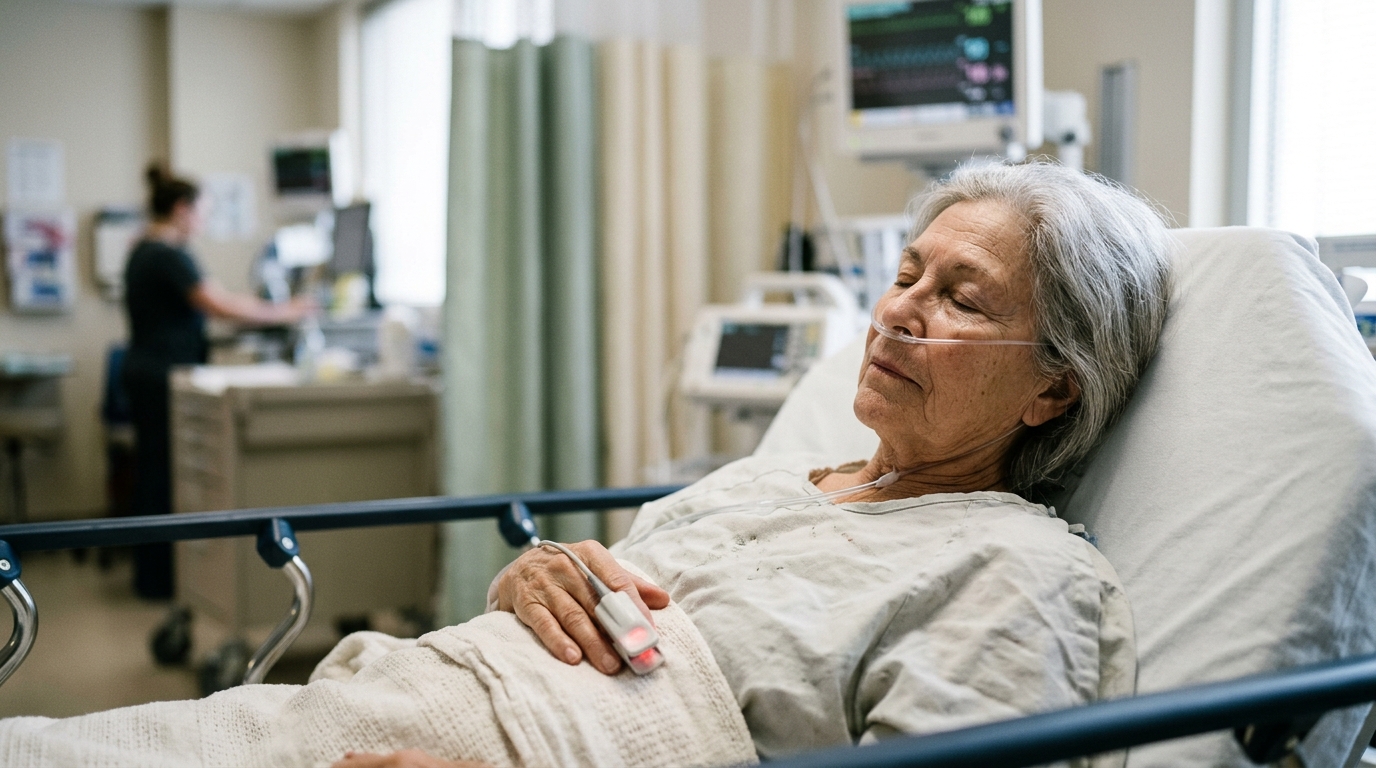
Older surgical patients routinely need less anaesthetic than younger ones, wake up more slowly, and run a higher risk of postoperative delirium and haemodynamic instability. For decades these observations were attributed mainly to the general functional decline of ageing.
The scale of the question is considerable. More than 300 million surgical procedures are performed worldwide each year, and a growing share involve older adults, a rapidly expanding demographic. Ageing reduces physiological reserve and is associated with heightened sensitivity to anaesthetic agents, reduced drug requirements and prolonged emergence. The review, written by anaesthesiology and clinical researchers at Hospital Clínico Universidad de Chile, sets out to separate what is well established from what remains hypothesis.
The authors begin with a distinction that matters clinically. Pharmacokinetics describes how a dose becomes a plasma or effect-site concentration; pharmacodynamics describes what that concentration does once it reaches its target. Ageing alters both. Reduced total body water and lean body mass, more adipose tissue, and declines in hepatic mass, hepatic blood flow, renal perfusion and glomerular filtration change drug distribution and clearance. Distribution into highly perfused tissues slows, and lipophilic drugs show larger apparent volumes of distribution and longer elimination half-lives. These changes explain why older adults need smaller bolus doses and lower maintenance infusion rates to reach plasma concentrations comparable to those of younger patients.
But pharmacokinetics alone cannot account for the full picture. At a given plasma or effect-site concentration, older adults often show more profound clinical and neurophysiological effects, indicating a genuine pharmacodynamic shift. The minimum alveolar concentration of volatile anaesthetics falls by roughly 6% per decade after age 40, and contemporary pharmacokinetic-pharmacodynamic models show age-dependent reductions in the effect-site concentrations of propofol and remifentanil needed to produce defined electroencephalographic effects. Proposed mechanisms include altered GABAA, NMDA and opioid receptor function, loss of neuronal synapses, disturbed intracellular calcium handling, cortical structural change and reduced functional connectivity. The review’s central proposal is that neuroimmune ageing may be one of the modifiers of this concentration-effect relationship itself.
The clinical signature of that shift is visible on the EEG. Processed indices such as the bispectral index and spectral entropy show that older patients may reach deeper anaesthetic states at lower drug concentrations. Burst suppression, an alternating pattern of high-voltage activity and electrical quiescence reflecting profound cortical depression, is particularly telling: it marks relative overdosage, a standard dose becoming disproportionately high for a vulnerable brain. Older patients show age-related increases in the duration and prevalence of intraoperative burst suppression, suggesting greater susceptibility to excessive cortical depression under standard dosing strategies.
The review then turns to the molecular substrate. Propofol, etomidate and volatile anaesthetics produce hypnosis through positive allosteric modulation of GABAA receptors, making that receptor a plausible pharmacodynamic target for age-related and inflammation-related change. Importantly, the inflammatory effects differed depending on the anaesthetic and the endpoint assessed, supporting the idea that neuroimmune modulation of anaesthetic sensitivity is agent- and circuit-specific rather than uniform.
Inflammaging itself arises from converging mechanisms including cellular senescence, immune cell reprogramming, gut dysbiosis and mitochondrial dysfunction, which together sustain a pro-inflammatory milieu. A hallmark feature is the persistent activation of monocytes and macrophages, which acquire pro-inflammatory phenotypes and elevate systemic levels of cytokines such as TNF-α, IL-1β and IL-6. Macrophages matter here because of their longevity, plasticity and tissue-resident sentry functions; during ageing they shift toward a primed, hyperresponsive state with exaggerated responses to minimal stimuli and impaired resolution of inflammation. They also contribute to the senescence-associated secretory phenotype, a cocktail of cytokines, chemokines and matrix-remodelling enzymes that perpetuates neuroinflammation.
How peripheral immune signals reach the brain is the review’s mechanistic core. The blood-brain barrier, once considered an immunoprivileged interface, becomes more permeable with ageing and systemic inflammation. Age-related endothelial changes, including tight junction disruption, altered transporter function and increased transcytosis, facilitate the passage of peripheral immune mediators into the central nervous system, and monocyte-derived macrophages can infiltrate the perivascular space and brain parenchyma. Yet macrophage-associated inflammation may also act without direct cellular entry: circulating cytokines, chemokines and complement components can be sensed by the cerebral endothelium, which the authors describe as an active component of the neurovascular unit rather than a passive barrier. Perivascular and border-associated macrophages provide a further interface between peripheral and central immunity. Separate work demonstrated that neuronal hyperactivity induces region-specific, C1q-dependent synapse loss in the adult hippocampus, linking complement activation to activity-dependent synaptic remodelling. Neither study examined anaesthetic sensitivity directly, but together they suggest complement dysregulation as an additional pathway connecting peripheral immune ageing with synaptic vulnerability.
Preclinical models supply the most direct evidence that inflammation changes anaesthetic depth. Proposed mechanisms include cytokine-mediated disruption of long-term potentiation and of glutamatergic and GABAergic signalling, primed aged microglia responding to systemic inflammatory signals with exaggerated cytokine production, and inflammatory alteration of ion channel function, oscillatory bursting and synaptic communication within thalamocortical circuits, which are critical for maintaining consciousness and are disrupted by general anaesthetics.
The clinical evidence, however, remains associative and incomplete. Most human data come from observational studies and anaesthetic-depth trials in older surgical patients, in whom low processed EEG values, intraoperative EEG suppression, or suppression at lower volatile anaesthetic concentrations have been linked to postoperative delirium and adverse neurocognitive outcomes. In the ENGAGES trial, EEG-guided anaesthesia reduced burst suppression duration but did not significantly affect delirium incidence, suggesting burst suppression is unlikely to be explained by dosage alone and may reflect broader biological vulnerability. A retrospective analysis of intraoperative EEG suppression in more than 1,000 patients found suppression duration to be a strong predictor of postoperative delirium independent of anaesthetic dose. Perioperative studies have also linked delirium with blood-brain barrier dysfunction and neuroinflammatory biomarkers in cerebrospinal fluid: a prospective cohort found perioperative changes in the CSF-to-plasma albumin ratio associated with delirium incidence and severity, alongside CSF IL-6 and lactate responses, while dynamic contrast-enhanced MRI showed increased hippocampal leakage in APOE4 carriers that correlated with cognitive decline. Elevated preoperative inflammatory markers predicted higher rates of delirium in 560 older adults undergoing major surgery, and postoperative cytokine surges were linked to delirium onset in 351 older adults. The authors are explicit that these associations do not establish that immune dysregulation directly alters anaesthetic pharmacodynamics in humans.
What would move the field forward? The review calls for prospective studies combining simultaneous immunological profiling, neurophysiological monitoring and pharmacokinetic-pharmacodynamic modelling, using biomarkers such as the CSF-to-plasma albumin ratio and EEG-derived indices of brain responsiveness. It also argues that chronological age alone inadequately captures biological heterogeneity: frailty, which reflects multidimensional loss of physiological reserve and is closely intertwined with immunosenescence and inflammaging, should be assessed alongside age, and sex-stratified analyses should account for reproductive ageing as a potential modifier of neuroimmune and anaesthetic phenotypes. Interventional probes, including perioperative anti-cytokine therapies, senolytics or strategies aimed at preserving barrier integrity, remain investigational, and immune biomarkers are not currently validated for anaesthetic dose selection. The hypothesis is testable, the authors conclude, and testing it will require bridging anaesthesiology, immunology, neurobiology and geriatrics in integrative perioperative studies.
Subject of Research: How inflammaging and neuroimmune ageing may alter anaesthetic sensitivity in older surgical patients
Article Title: Ageing and anaesthetic sensitivity: the role of inflammaging in the vulnerable surgical brain
Article References: Díaz-Papapietro, C., Ríos, D., Brisso, R., Gutiérrez, R., Egaña, J. I., & Penna, A. (2026). Ageing and anaesthetic sensitivity: the role of inflammaging in the vulnerable surgical brain. Frontiers in Medicine, 13, Article 1920909. https://doi.org/10.3389/fmed.2026.1920909
Image Credits: AI Generated
DOI: 10.3389/fmed.2026.1920909
Keywords: anaesthesia, ageing, inflammaging, macrophages, blood-brain barrier, neuroinflammation, EEG, burst suppression, pharmacodynamics, postoperative delirium, GABAA receptor, frailty
News Source: Cassandra Pierce. (November 6, 2026). Inflammaging may make the ageing brain unusually sensitive to anaesthesia. Scienmag.