A new bispecific antibody designed to drag immune cells into CLDN18.2-positive tumors has shown dose-dependent tumor suppression, precise tumor targeting, and no systemic immunotoxicity in preclinical testing, according to a study published in Drugs in R&D. The molecule, called AHT-102, is a Fc-free CLDN18.2 × CD3 bispecific antibody built on the XFab platform, and its developers at Beijing Immunoah Pharma Tech and collaborating institutions report that it achieved a maximum tolerated dose 136.5 times higher than the projected clinical starting dose in humanized mice. The findings, published on 26 February 2026, provide what the authors describe as a robust scientific rationale for moving the compound into clinical trials in patients with CLDN18.2-positive cancers.
AHT-102 was engineered specifically to address these limitations through two structural choices: the removal of the Fc region and the placement of a low-affinity CD3-binding module at the C-terminus of the molecule.
The Fc-free design is central to the safety strategy. Prior research cited by the authors also suggests that intact Fc domains may sequester T cells in the periphery, hindering their infiltration into subcutaneous solid tumors. The smaller size, however, may facilitate superior tumor tissue penetration, and several TCE molecules built on the same XFab platform have already progressed into clinical trials.
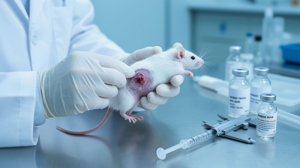
To test anti-tumor efficacy, researchers used a subcutaneous xenograft model in which human gastric cancer NUGC4-CLDN18.2 cells were implanted into NSG mice that then received human peripheral blood mononuclear cells (PBMCs) to create a humanized immune system. Once tumors reached an average volume of approximately 53 cubic millimeters, sixty mice were randomized into four groups of ten: a vehicle control and three AHT-102 dose groups at 0.1, 0.3, and 1.0 mg/kg. Treatment was given intravenously once daily for three days, followed by a three-day break, across four cycles. By the end of the study on post-graft day 21, tumor growth inhibition rates based on volume were 16 percent, 34 percent, and 47 percent for the low, medium, and high doses respectively, and all treated groups showed significantly lower tumor volumes than the vehicle group (p < 0.01). Tumor weight inhibition reached 51 percent at the highest dose, with all treatment groups showing significantly reduced tumor weights compared with controls (p < 0.01). No animal mortality occurred in any group, and the authors attribute a downward trend in body weight across all cohorts, including the vehicle group, to graft-versus-host disease induced by the human PBMCs rather than drug-specific toxicity.
Pharmacokinetic analysis in human CD3EDG transgenic mice, which carry humanized CD3ε, CD3δ, and CD3γ genes on a C57BL/6J background, revealed linear characteristics across a dose range of 0.1 to 2.5 mg/kg. After single intravenous injections, maximum concentration ratios of 1:4.76:25.1 and area-under-the-curve ratios of 1:5.18:26.4 closely tracked the dose ratios of 1:5:25. Refined terminal half-lives were 0.762 hours, 3.05 hours, and 4.25 hours for the 0.1, 0.5, and 2.5 mg/kg doses respectively, with similar clearance rates of 78.2 to 81.8 mL/h/kg across all groups. The apparent volume of distribution, ranging from 89.9 to 705 mL/kg, exceeded the plasma volume of mice, suggesting that AHT-102 distributes outside the blood vessels.
Tissue distribution studies used radiolabeled AHT-102 in hCD3EDG mouse colorectal cancer models bearing MC38-hCLDN18.2 tumors. Following a single intravenous dose of iodine-125-labeled AHT-102 at 2.5 mg/kg, exposure levels across tissues ranked from highest to lowest as serum, kidney, lung, stomach, liver, tumor, spleen, adrenal gland, heart, esophagus, duodenum, femur, ovaries, bladder, intestinal contents, thymus, pancreas, testes, skeletal muscle, mesenteric lymph node, gallbladder, body fat, eyeball, and brain. In blood-rich tissues such as kidney, lung, and liver, concentrations peaked at two minutes and declined rapidly, mirroring serum. In contrast, the CLDN18.2-positive stomach and tumor tissues showed active sequestration, reaching peak concentrations at four hours post-dosing, significantly later than in the circulation, which the authors interpret as evidence of specific binding and accumulation.
Quantitative targeting metrics reinforced this interpretation. The tumor-to-blood ratio increased progressively from 0.01 at two minutes to 3.09 at twelve hours, representing a 300-fold enrichment of the drug within the tumor relative to serum over that window. At the twelve-hour terminal point, tumor concentration reached 495 equivalent nanograms per gram, substantially higher than the heart at 32.8 and skeletal muscle at 32.6, yielding a tumor-to-heart ratio of 15.1 and a tumor-to-muscle ratio exceeding 15. Minimal distribution in the brain indicated no penetration of the blood-brain barrier. Importantly, exposure in the spleen and mesenteric lymph nodes was lower than in the tumor, suggesting that AHT-102 bypassed the so-called T-cell sink, in which high-affinity CD3 binding sequesters drug in T-cell-rich secondary lymphoid organs rather than allowing it to reach target-positive tumors.
In vitro testing addressed the risk of nonspecific immune activation. When AHT-102 at concentrations from 0.0001 to 10 micrograms per milliliter was co-cultured with PBMCs from three healthy donors for 2, 4, 24, and 48 hours in the absence of CLDN18.2-positive cells, levels of interleukin-2, interleukin-4, interleukin-6, interleukin-8, interleukin-10, interferon-gamma, and tumor necrosis factor-alpha did not increase with either incubation time or drug concentration. This indicates that without target cells present, the antibody does not induce cytokine release from PBMCs, a finding consistent with the design goal of preventing systemic T-cell activation in the absence of tumor antigen engagement.
Safety was further evaluated in a five-week repeat-dose toxicity study involving 654 healthy hCD3EDG mice, evenly divided between males and females and assigned to vehicle control or AHT-102 doses of 1.50, 4.50, and 13.65 mg/kg given intravenously once weekly, followed by a six-week recovery period. The maximum tolerated dose was established at 13.65 mg/kg, at which the average area under the curve from zero to 168 hours after the final administration reached 236 hours times micrograms per milliliter, with a mean peak concentration of 158 micrograms per milliliter. No drug-related mortality or severe morbidity was recorded, and comprehensive monitoring of general observations, food consumption, body weight, gross pathology, ophthalmic examinations, hematology, coagulation, and urinalysis revealed no drug-related abnormalities. Blood biochemistry showed dose-dependent decreases in albumin and total protein in female mice from the mid- and high-dose groups and reductions in albumin and albumin/globulin ratios in males, but these changes remained within normal ranges and resolved by the end of the recovery period.
Histopathological examination did identify adverse changes in stomach tissue, including mild to severe enlargement of mucous cells, regenerative hyperplasia of the mucosa, single-cell necrosis, neutrophil infiltration, and reactive squamous epithelial hyperplasia, with mild focal erosion or ulceration in female mice from the mid- and high-dose groups and males from the high-dose group. The authors characterize this as an expected on-target effect arising from endogenous CLDN18.2 expression in normal gastric tissue, a phenomenon also observed with the approved CLDN18.2-targeted antibody zolbetuximab. All pathological alterations had fully resolved by the end of the recovery period, indicating reversibility upon drug discontinuation. The authors note that direct immunohistochemical evidence of T-cell infiltration into tumors was not collected, so the observed dose-dependent tumor suppression serves as functional but indirect validation of T-cell recruitment. They also point to potential combination strategies, noting that phase III SPOTLIGHT and GLOW trials established zolbetuximab plus chemotherapy as a standard of care for previously untreated CLDN18.2-positive gastric or gastroesophageal junction adenocarcinoma, and that pairing T-cell engagers with immune checkpoint inhibitors could be particularly compelling for patients with microsatellite-stable tumors who traditionally respond poorly to checkpoint blockade alone. These are preclinical findings in mouse models, and clinical safety and efficacy in humans remain to be established.
Subject of Research: Preclinical evaluation of a Fc-free CLDN18.2 × CD3 bispecific antibody for CLDN18.2-positive cancers
Article Title: Preclinical Evaluation of AHT-102, a CLDN18.2 × CD3 Bispecific Antibody: Pharmacokinetics, Anti-Tumor Efficacy, Tissue Distribution, and Safety Profile
Article References: Chu, H., Xu, G., Liu, Y., Qian, N., Li, Y., Liu, Y., Pan, X., Gao, X., Ou, L., Lv, J., & Song, H. (2026). Preclinical Evaluation of AHT-102, a CLDN18.2 × CD3 Bispecific Antibody: Pharmacokinetics, Anti-Tumor Efficacy, Tissue Distribution, and Safety Profile. Drugs in R&D, 26(1), 39-50. https://doi.org/10.1007/s40268-026-00535-y
Image Credits: AI Generated
DOI: 10.1007/s40268-026-00535-y
Keywords: AHT-102, CLDN18.2, CD3 bispecific antibody, T-cell engager, gastric cancer, preclinical study, pharmacokinetics, tissue distribution, cytokine release syndrome, Fc-free antibody, tumor growth inhibition, humanized mouse model
News Source: Nathaniel Bowman. (October 30, 2026). Fc-Free CLDN18.2 × CD3 Antibody AHT-102 Shrinks Gastric Tumors in Mice. Scienmag.