Cardiovascular diseases remain the leading cause of global mortality, claiming roughly 20 million lives each year, and a newly published review argues that the next leap in treatment may come not from new drugs but from smarter delivery vehicles. Writing in the journal Discover Pharmaceutical Sciences, researchers Rufaida Wasim and Aditya Singh survey the rapidly growing field of engineered exosome-based nanocarriers, naturally derived vesicles that could ferry therapeutic cargo directly to diseased heart and vascular tissue while sparing healthy cells. The review, published as an open-access article on 3 July 2026, synthesizes preclinical evidence across myocardial infarction, atherosclerosis, heart failure, cardiac fibrosis and vascular inflammation, and concludes that exosomes represent a transformative strategy for cardiovascular drug delivery, provided that formidable manufacturing and regulatory barriers can be overcome.
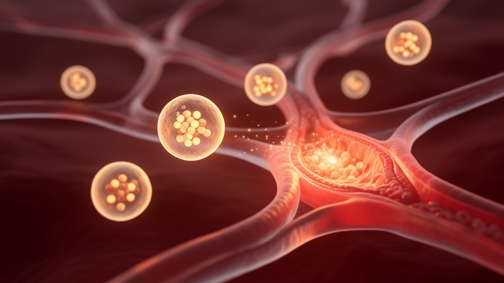
Exosomes are nanosized extracellular vesicles, typically 30 to 150 nanometers in diameter, that cells secrete constantly as part of intercellular communication. They arise from the endosomal pathway: the cell membrane buds inward to form multivesicular bodies, which then fuse with the plasma membrane and release their internal vesicles into the extracellular space. Their cargo is not random. RNA-binding proteins, lipid interactions and sequence motifs guide the selective sorting of proteins, lipids, mRNA, microRNA and other nucleic acids into each vesicle, meaning that an exosome’s contents reflect the physiological or pathological state of the cell that produced it. In the cardiovascular system, cardiomyocytes under ischemic stress release exosomes enriched with stress-responsive microRNAs such as miR-21 and miR-210, while endothelial cell-derived exosomes help regulate vascular inflammation and nitric oxide homeostasis.
What makes exosomes attractive as drug carriers is a combination of properties that synthetic nanoparticles struggle to match. Because their membranes closely resemble those of the parent cell, they enjoy intrinsic biocompatibility, low immunogenicity, prolonged circulation and a degree of natural tissue targeting through surface proteins such as integrins and adhesion molecules. In ischemic myocardium, for example, increased expression of adhesion molecules like ICAM-1 and VCAM-1 facilitates enhanced uptake of circulating exosomes, effectively exploiting the inflammatory microenvironment to concentrate the vesicles at the diseased site. Synthetic carriers such as lipid nanoparticles, polymeric nanoparticles, dendrimers and micelles have shown improved pharmacokinetics and controlled release in preclinical models, but they face immunogenicity, complement activation, rapid clearance by the mononuclear phagocyte system and long-term toxicity concerns, and they generally require extensive surface modification to achieve any targeting at all.
Native exosomes, however, are not clinical-grade delivery vehicles on their own, and much of the review is devoted to the engineering strategies that convert them into one. One widely used approach is pre-loading engineering: donor cells are genetically or biochemically modified before exosome isolation, so that therapeutic proteins or RNAs are packaged into the secreted vesicles through endogenous sorting. Overexpressing cardioprotective microRNAs in mesenchymal stem cells, for instance, enriches them within exosomes and improves myocardial repair outcomes in ischemic models. Surface targeting can be engineered the same way, by fusing exosomal membrane proteins such as Lamp2b with targeting peptides that direct the vesicles toward specific tissues, including ischemic myocardium. Alternatively, purified exosomes can be modified after isolation using click chemistry, covalent bonding or lipid insertion to attach ligands, antibodies or peptides; incorporating ischemia-targeting peptides or integrin-binding motifs can significantly increase accumulation within infarcted myocardium, although excessive chemical manipulation risks altering membrane integrity or inducing aggregation.
Getting cargo into the vesicle is its own engineering problem, and the review catalogs the trade-offs in detail. Passive loading, in which purified exosomes are simply incubated with hydrophobic drugs that diffuse across the lipid bilayer, is straightforward and preserves vesicle structure but yields low encapsulation efficiency, typically around 5 to 20 percent. Active methods transiently disrupt the membrane to improve loading: electroporation applies electrical pulses to admit nucleic acids such as siRNA or miRNA, achieving roughly 15 to 40 percent encapsulation for RNA cargo but risking nucleic acid aggregation and vesicle instability; sonication uses ultrasonic shear forces and reaches about 20 to 45 percent, particularly for hydrophobic drugs, but can damage membrane proteins needed for targeting; extrusion and freeze-thaw cycles achieve around 25 to 50 percent at the cost of altered vesicle morphology. Detergent-assisted loading with Triton X-100, which transiently solubilizes the lipid bilayer before the membrane reforms, has reported intermediate efficiencies of roughly 30 to 60 percent, though residual detergent toxicity and structural alterations remain concerns for clinical translation.
By comparison, the synthetic competition sets a high bar on loading capacity alone. Liposomes typically achieve drug loading efficiencies of approximately 40 to 90 percent and polymeric nanoparticles 50 to 95 percent, depending on formulation, whereas exosomes generally fall in the 10 to 50 percent range depending on method. The authors argue, however, that absolute loading percentage is not the decisive metric: exosomes compensate with superior targeting efficiency and enhanced cellular uptake, potentially reducing the therapeutic dose required. Optimization of the loading-to-function ratio, they contend, matters more than raw encapsulation figures, and post-loading characterization, including particle size analysis, zeta potential measurement, transmission electron microscopy and cargo quantification, is essential to ensure structural integrity and reproducibility.
The preclinical evidence base spans several disease models and molecular pathways. In myocardial infarction, engineered exosomes loaded with cardioprotective microRNAs such as miR-21, miR-126 and miR-210 have shown enhanced angiogenesis, reduced apoptosis and improved cardiac function in animal models; mechanistically, miR-21-mediated inhibition of PTEN activates the PI3K/Akt survival cascade, while miR-210 enhances hypoxia adaptation and mitochondrial function. Mesenchymal stem cell-derived exosomes enriched with miR-126 have promoted endothelial proliferation and neovascularization in ischemic myocardium by activating the VEGF pathway. In atherosclerosis, delivery of anti-miR-33 via engineered exosomes promotes cholesterol efflux by upregulating the transporter ABCA1 in macrophages, reducing plaque burden, while exosomes carrying anti-inflammatory molecules or siRNAs modulate macrophage polarization within plaques. In cardiac fibrosis models, CRISPR/Cas9-loaded exosomes targeting TGF-β signaling have demonstrated reduced fibroblast activation and decreased collagen deposition. Exosomes have also carried statins for plaque stabilization, siRNA against apoptosis genes such as Bax and caspase-3 after infarction, antioxidants like curcumin and resveratrol, and pro-angiogenic proteins such as VEGF.
The pharmacokinetic picture is more nuanced than for synthetic carriers. Exosomes are administered intravenously, intracoronarily or by intramyocardial injection in cardiovascular applications; intravenous delivery is minimally invasive but leads to rapid uptake by the mononuclear phagocyte system, particularly in the liver and spleen, whereas local routes enhance regional bioavailability and tissue retention. Biodistribution studies with fluorescent and radiolabeled exosomes show preferential accumulation in liver, spleen, lungs and injured tissues, with inflamed or ischemic cardiac tissue taking up more vesicles due to increased vascular permeability. Native exosomes display short systemic half-lives ranging from minutes to a few hours, prompting exploration of PEGylation and membrane modification to prolong circulation. They are cleared mainly by macrophage-mediated phagocytosis in the liver and spleen and degraded in lysosomes, and their biodegradable lipid and protein composition reduces the long-term tissue accumulation concerns associated with certain polymeric nanoparticles. Notably, exosomal membranes themselves contain bioactive proteins and lipids that may synergistically enhance cardioprotection, meaning pharmacokinetic-pharmacodynamic relationships differ from conventional nanocarriers and require specialized modeling.
Despite the encouraging experimental record, the review is emphatic that clinical translation remains in its early stages and that most evidence supporting exosome-mediated cardioprotection derives from animal models. Scalable manufacturing is a major barrier: conventional ultracentrifugation is labor-intensive and yields heterogeneous vesicle populations, and while tangential flow filtration and microfluidic platforms offer improved scalability, consistent batch-to-batch reproducibility remains difficult. Exosome heterogeneity, donor cell variability and differing purity profiles across isolation methods, from size-exclusion chromatography to polymer precipitation to immunoaffinity capture, complicate standardization. Safety questions persist around off-target effects, unintended immune modulation, horizontal gene transfer, hepatic and splenic accumulation, and the potential transmission of oncogenic or pathogenic material from donor cells, which necessitates rigorous donor screening. Regulatory classification is itself unsettled: depending on jurisdiction and modification, exosome products may be treated as biological medicinal products or advanced therapy medicinal products, each with distinct approval pathways requiring defined source cell characterization, GMP-compliant manufacturing, potency assays, biodistribution and toxicity data, and batch consistency validation.
Early-phase clinical trials of mesenchymal stem cell-derived exosomes in inflammatory and regenerative conditions have demonstrated favorable safety profiles, but cardiovascular-specific exosome trials remain limited and exploratory, and preclinical large-animal studies showing improvements in left ventricular ejection fraction, infarct size and angiogenesis have yet to be matched by randomized human trials. The authors argue that bridging this gap will require robust pharmacokinetic, pharmacodynamic and dose-optimization studies, well-designed multicenter clinical trials, and interdisciplinary collaboration among pharmacologists, bioengineers, clinicians and regulatory authorities. Looking forward, they point to hybrid biomimetic nanoparticles, artificial exosome mimetics, stimulus-responsive programmable vesicles, integration with CRISPR-based gene editing, and artificial intelligence-guided engineering as directions that could sharpen therapeutic precision. If standardization initiatives and GMP-compliant production mature as anticipated, engineered exosome nanocarriers may yet move from laboratory promise to a genuine cell-free alternative that preserves the paracrine benefits of stem cell therapy, without the immune rejection, tumorigenicity and poor cell survival risks that have long shadowed cell transplantation in cardiac medicine.
Subject of Research: Engineered exosome-based nanocarriers for targeted drug delivery in cardiovascular diseases
Article Title: Engineered exosome nanocarriers for targeted cardiovascular therapy
Article References: Wasim, R., & Singh, A. (2026). Engineered exosome nanocarriers for targeted cardiovascular therapy. Discover Pharmaceutical Sciences, 2(1), Article 14. https://doi.org/10.1007/s44395-026-00048-2
Image Credits: AI Generated
DOI: 10.1007/s44395-026-00048-2
Keywords: exosomes, nanocarriers, cardiovascular disease, drug delivery, myocardial infarction, atherosclerosis, cardiac fibrosis, microRNA, CRISPR, preclinical research, targeted therapy, drug loading
News Source: Louis Brooks. (October 29, 2026). Engineered exosome nanocarriers emerge as targeted cardiovascular drug delivery platform. Scienmag.