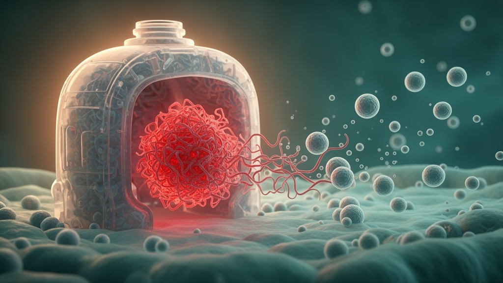
When a cell’s waste-disposal systems fail, the damage does not necessarily stay contained. A new open-access study published in Cell Communication and Signaling on 28 October 2026 reports that shutting down the two major protein-degradation pathways of the cell — the ubiquitin–proteasome system (UPS) and the autophagy–lysosome pathway (ALP) — dramatically changes what cells package into the tiny membrane bubbles known as extracellular vesicles (EVs), and that these altered vesicles can transmit proteotoxic stress to healthy recipient cells.
The research, a collaboration led by Elena Casarotto, Daisy Sproviero, Letizia Messa and colleagues under the senior authorship of Angelo Poletti and Valeria Crippa, focused on protein quality control (PQC), the coordinated network of chaperone proteins, the UPS and the ALP that normally keeps the proteome soluble and functional. Disruption of this network is a hallmark of TDP-43–associated neurodegenerative diseases, including amyotrophic lateral sclerosis (ALS) and frontotemporal dementia (FTD), conditions in which the RNA-binding protein TDP-43 misfolds, fragments and aggregates inside affected neurons. Because EVs are known to ferry misfolded proteins out of cells, the team asked how PQC failure reshapes the cargo of these vesicles and whether the result contributes to disease spread.
To find out, the researchers cultured motor neuron–like NSC-34 cells and isolated two EV populations under standard differential-centrifugation conventions: larger vesicles from a 20,000 × g pellet (20K EVs) and smaller vesicles from a 100,000 × g pellet (100K EVs). Vesicles were collected both under basal conditions and after pharmacological inhibition of either the UPS or the ALP. The team then characterized the vesicles with nanoparticle tracking analysis, electron and super-resolution microscopy, biochemical assays and biophysical spectroscopies, profiled their miRNA cargo by small RNA sequencing, and tested the functional effects of the vesicles on recipient cells.
The results show that PQC impairment markedly reshapes EV cargo. Both UPS and ALP inhibition enhanced the extracellular release of misfolded TDP-43 C-terminal fragments, the autophagy receptor SQSTM1/p62 and ubiquitinated proteins — a molecular signature of a cell struggling to clear damaged proteins. Notably, blocking the ALP specifically increased the EV-associated export of insoluble phosphorylated TDP-43 at the disease-relevant Ser409/410 residues, together with MAP1LC3, a core autophagy protein, suggesting that stalled autophagic intermediates are being shunted into the vesicle route.
The biophysical signatures of the exported aggregates differed strikingly between the two inhibition conditions. UPS inhibition favored the secretion of compact, large aggregates rich in β-sheet structure — the tightly ordered packing typical of mature amyloid-like material. ALP inhibition, by contrast, promoted the release of smaller aggregated species with increased surface hydrophobicity, a profile consistent with intermediate aggregates that have not yet matured into compact fibrils. In other words, which degradation pathway fails appears to determine not just how much misfolded protein leaves the cell, but in what aggregation state it travels.
PQC dysfunction also remodeled the vesicles’ regulatory cargo. The most pronounced changes appeared in the small 100K EVs, whose miRNA content was enriched — after PQC disruption — for pathways linked to endoplasmic reticulum (ER) stress and prion-related processes. This indicates that vesicles released by stressed cells carry not only proteinaceous seeds but also a nucleic-acid payload biased toward stress and misfolding programs.
The functional experiments drove the point home. When EVs generated under conditions of PQC impairment were applied to recipient cells, they reduced cell viability and triggered proteotoxic responses connected to ER stress and prion-related pathways. The vesicles also activated Notch1–c-Myc signaling and switched on transcriptional programs associated with cell death. Together, these findings support the authors’ conclusion that EV secretion acts as a protective, regulated response to proteostasis imbalance — a way for a distressed cell to offload toxic material — but that this escape route carries an undesired consequence: the propagation of non–cell-autonomous proteotoxicity in TDP-43 proteinopathies.
The study’s scope should be kept in perspective. All experiments were performed in a motor neuron–like cell line under acute pharmacological inhibition of the degradation pathways, so the findings establish a mechanism in a controlled cellular model rather than in patient tissue or living organisms. The work was shared early as a peer-reviewed, accepted manuscript carrying a permanent DOI, and the published version remains subject to final editorial updates. Even so, the results offer a concrete mechanistic picture of how the same proteostasis failure that kills a neuron may also equip its vesicles to spread the damage — a concept that could inform future strategies aiming to intercept EV-mediated spread in ALS and FTD.
Subject of Research: How impaired protein quality control reshapes extracellular vesicle cargo and propagates proteotoxicity in TDP-43 proteinopathies
Article Title: Proteasome and autophagy inhibition reshapes extracellular vesicle cargo and spreads proteotoxicity
Article References: Casarotto, E., Sproviero, D., Messa, L., Brivio, A., Conti, A. C., Gagliani, M. C., Cornaggia, L., Cozzi, M., Chierichetti, M., Marchesi, V., Mohamed, A., Piccolella, M., Ferrari, V., Tedesco, B., Cristofani, R., Rusmini, P., Galbiati, M., Carelli, S., Cortese, K., … Crippa, V. (2026). Proteasome and autophagy inhibition reshapes extracellular vesicle cargo and spreads proteotoxicity. Cell Communication and Signaling. https://doi.org/10.1186/s12964-026-03327-8
Image Credits: AI Generated
DOI: 10.1186/s12964-026-03327-8
Keywords: extracellular vesicles, TDP-43, proteasome, autophagy, protein quality control, ALS, frontotemporal dementia, proteostasis, miRNA, ER stress, prion-like propagation, NSC-34 cells
News Source: Diana Fleming. (October 28, 2026). Blocked protein cleanup reroutes toxic TDP-43 cargo through extracellular vesicles. Scienmag.