Chronic alcohol drinking damages the heart muscle in ways that have long been attributed to oxidative stress, inflammation and metabolic disruption, but a new experimental study points to a more specific molecular culprit: a copper-triggered form of regulated cell death called cuproptosis. In a paper published in the Journal of Cellular and Molecular Medicine, researchers at Wuhan University report that a gene involved in branched-chain amino acid metabolism, DBT, behaves like a switch in this process. When its activity falls in heart cells, copper accumulates and cuproptosis-related injury follows; when it is boosted experimentally, the damage recedes.
The team built alcoholic cardiomyopathy in mice by feeding them an ethanol-containing Lieber-DeCarli liquid diet for ten weeks, ramping the ethanol concentration gradually from 1 percent to 5 percent during an adaptive period and then stepping it up every two weeks until it reached 5.4 percent. Thirty male C57BL/6 mice were randomly assigned, eighteen to ethanol feeding and twelve to an isocaloric ethanol-free control diet in which maltodextrin replaced the alcohol calories. The ethanol-fed animals gained weight more slowly and weighed significantly less than controls from week six onward. By the end of the protocol, their blood showed elevated alanine aminotransferase, aspartate aminotransferase, blood alcohol and B-type natriuretic peptide, a combination the authors read as successful alcohol exposure with cardiac injury.
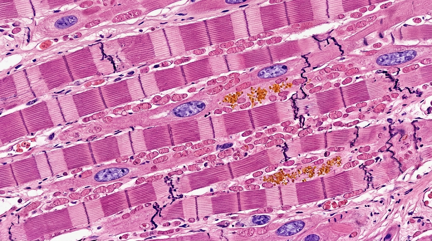
Echocardiography after ten weeks confirmed that the model had taken hold. Compared with controls, the ethanol group displayed chamber dilation and impaired contractile function, with significantly reduced left ventricular ejection fraction and fractional shortening and markedly increased internal diameters in both diastole and systole. Tissue Doppler measurements revealed a reduced early diastolic mitral annular velocity and an increased E/e′ ratio, indicating diastolic dysfunction as well. Histology showed disorganized myocardial architecture, enlarged ventricles, thinned walls and inflammatory infiltration, while Masson’s trichrome staining revealed extensive interstitial collagen deposition, the hallmark of fibrosis. Morphometrically, the alcohol group had lower body weight, higher heart weight and an elevated heart-weight-to-body-weight ratio.
With the model established, the researchers turned to transcriptome sequencing of left ventricular tissue from four control and four ACM mice. Applying exploratory screening criteria of an absolute log-fold change above 0.2 and a nominal p value below 0.05, they identified 2485 candidate genes. Intersecting these with a published list of 86 cuproptosis-related genes yielded seven overlapping candidates: VEGFA, CP, SQSTM1, SLC31A2, DBT, PDK1 and SERPINE1. Only VEGFA and CP survived Benjamini-Hochberg correction for multiple testing; the rest, including DBT, remained exploratory, hypothesis-generating candidates rather than statistically significant differentially expressed genes after false discovery rate control.
Among the seven, the authors selected DBT, CP and PDK1 for laboratory validation because violin plots showed nominal p values below 0.05 for all three. Quantitative PCR and Western blotting then narrowed the field further: only DBT differed significantly between control and ACM hearts at both the mRNA and protein levels. DBT was consistently downregulated in two independent RNA-sequencing cohorts, with a log-fold change of roughly −0.32 in each, but it did not reach false discovery rate significance in either. The authors are explicit that DBT is therefore a reproducible exploratory candidate, not a conventionally significant hit. Gene set enrichment analysis linked DBT expression to several cardiomyopathy-related pathways, including dilated and hypertrophic cardiomyopathy, arrhythmogenic right ventricular cardiomyopathy and viral myocarditis, and gene set variation analysis suggested an association with branched-chain amino acid catabolic pathways.
The copper connection came next. Serum copper was significantly elevated in the ACM mice, and when the team isolated adult cardiomyocytes from the animals’ hearts, intracellular copper content was also significantly increased, indicating genuine copper accumulation inside the muscle cells rather than merely in the blood. The molecular signature of cuproptosis accompanied this burden: FDX1 and the copper transporter SLC31A1 were decreased, the chaperone HSP70 was increased, and Western blotting plus immunofluorescence showed increased oligomerization and aggregation of DLAT, a lipoylated mitochondrial protein whose clumping is a defining event in cuproptosis. During this cell death process, copper binds directly to lipoylated proteins in the tricarboxylic acid cycle, triggering proteotoxic stress, loss of iron-sulfur cluster proteins and ultimately cell death, a mechanism distinct from apoptosis, necrosis, pyroptosis and ferroptosis.
To test whether DBT downregulation is a driver rather than a bystander, the researchers manipulated the gene in AC16 human cardiomyocyte-like cells. Silencing DBT reduced cell viability, induced morphological damage and reproduced the cuproptosis-related molecular changes seen in the mouse hearts, along with increased intracellular copper. Mitochondrial function deteriorated in parallel: reactive oxygen species rose, ATP fell, the NAD+/NADH ratio dropped and mitochondrial membrane potential was lost. Strikingly, tetrathiomolybdate, a copper chelator, markedly alleviated these injuries, whereas MitoQ, a mitochondria-targeted antioxidant, partially restored membrane potential but failed to significantly improve the other defects. That pattern, the authors argue, supports the copper-dependent nature of DBT knockdown-induced injury, since an antioxidant could not rescue what a copper chelator could.
The reciprocal experiment strengthened the case. Exposing AC16 cells to 500 millimolar ethanol for 24 hours reduced viability, raised intracellular copper and induced the same cuproptosis-related molecular alterations and mitochondrial dysfunction observed in the mouse model. Overexpressing DBT in these ethanol-treated cells alleviated the copper accumulation, the molecular injury and the mitochondrial dysfunction, performing similarly to the copper chelator TTM. In contrast, inhibitors targeting apoptosis, ferroptosis and autophagy-related processes, namely Z-VAD, Ferrostatin-1 and 3-methyladenine, did not produce statistically significant improvements under the treatment conditions and sample sizes used, though the authors caution that these negative results do not exclude those alternative cell death pathways.
The mechanistic story the authors propose runs through metabolism. DBT encodes the E2 subunit, dihydrolipoamide branched-chain transacylase, of the branched-chain α-keto acid dehydrogenase complex, which mediates branched-chain amino acid catabolism. Because branched-chain amino acid catabolites feed into the TCA cycle, and because cuproptosis hinges on lipoylated TCA-cycle proteins, impaired BCKDH-dependent metabolism could plausibly alter a cell’s susceptibility to copper-triggered death. Reduced DBT expression has previously been associated with dilated cardiomyopathy and heart failure, and the new findings suggest DBT may link mitochondrial metabolic dysfunction to cuproptosis-related cardiomyocyte injury. The authors stress, however, that this metabolic link remains hypothetical: they did not measure BCKDH activity, branched-chain amino acid flux, mitochondrial respiration or TCA-cycle activity, and their data do not establish that copper directly induces DBT degradation or that DBT buffers intracellular copper.
Several limitations frame how far the conclusions travel. No cardiomyocyte-specific DBT overexpression or knockout models were created, so the in vivo data support associations among DBT downregulation, copper accumulation, cuproptosis-related alterations and alcoholic cardiomyopathy, but do not establish a causal role for cuproptosis in cardiac dysfunction or disease progression in living animals. Only male mice were studied, leaving sex differences unexplored, and some mechanistic experiments included as few as three independent biological replicates without a prospective power calculation. AC16 cells, moreover, do not fully recapitulate primary human cardiomyocytes or the complexity of human disease, and interspecies differences in cardiac mitochondrial metabolism may affect translation. Within those boundaries, the study offers a coherent cellular-level argument: in this experimental system, DBT downregulation promotes copper-dependent, cuproptosis-related cardiomyocyte injury, DBT overexpression and copper chelation both blunt ethanol-induced damage, and whether DBT regulates alcoholic cardiomyopathy through cuproptosis in vivo remains an open question for future work with cardiomyocyte-specific manipulation and in vivo rescue experiments.
Subject of Research: Copper-dependent cuproptosis-related injury and the DBT gene in experimental alcoholic cardiomyopathy
Article Title: DBT Modulates Copper‐Dependent Cuproptosis‐Related Injury in Experimental Alcoholic Cardiomyopathy
Article References: Pan, Z., Gao, L., Ling, Y., & Cen, X. (2026). DBT Modulates Copper‐Dependent Cuproptosis‐Related Injury in Experimental Alcoholic Cardiomyopathy. Journal of Cellular and Molecular Medicine, 30(21), Article e71421. https://doi.org/10.1111/jcmm.71421
Image Credits: AI Generated
DOI: 10.1111/jcmm.71421
Keywords: alcoholic cardiomyopathy, cuproptosis, copper, DBT, mitochondrial dysfunction, cardiomyocytes, DLAT aggregation, Lieber-DeCarli diet, AC16 cells, copper chelation, RNA sequencing, branched-chain amino acid metabolism
News Source: Daisy Hatcher. (November 6, 2026). Alcohol-Linked Heart Damage in Mice Tied to Copper-Driven Cell Death Pathway. Scienmag.