Prostate cancer remains one of the most frequently diagnosed malignancies and a leading contributor to cancer mortality among men globally, ranking second in overall incidence and fifth in cancer death according to worldwide epidemiological evaluations. For four decades, prostate-specific antigen testing has functioned as the standard frontline screening tool, yet its low diagnostic specificity, high false-positive rate, and modest sensitivity for early localized disease frequently postpone accurate detection until tumors have reached advanced or metastatic stages. Even following primary intervention, a substantial fraction of patients undergoing initial androgen deprivation therapy eventually transition toward metastatic castration-resistant prostate cancer, a lethal disease state that lacks definitive curative interventions. Facing these persistent clinical hurdles, extracellular vesicles known as exosomes have emerged as indispensable non-invasive biological windows into tumor behavior. Protected by an enduring phospholipid bilayer that prevents the proteolytic and nucleolytic degradation typical of unencapsulated serum components, exosomal cargoes maintain remarkable biochemical stability in peripheral biofluids such as blood, urine, and seminal plasma. Among these encapsulated molecules, exosomal proteins represent the most immediately actionable candidates for clinical laboratory assays, providing directly measurable indicators capable of mapping cellular signaling networks across distinct stages of oncogenic development.
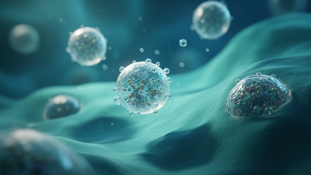
Exosomes are small vesicles of endosomal origin, typically spanning diameters between 30 and 150 nanometers, that are generated through the fusion of multivesicular bodies with the cell membrane. Their biogenesis relies on endosomal sorting complexes required for transport machinery alongside endosomal sorting complex-independent pathways, including the well-characterized syndecan-syntenin-ALIX molecular route. Far from being an accidental or passive sampling of the cytoplasm, the internal protein cargo within these nanometer-scale vesicles is sorted via deliberate biological processes that involve post-translational ubiquitination, association with specialized lipid microdomains, and interactions within tetraspanin-enriched membrane platforms. Once secreted into the extracellular space, exosomes engage neighboring or distant target tissues through direct surface receptor interactions, complete membrane fusion, or receptor-mediated phagocytosis, delivering molecular messages powerful enough to reprogram recipient cell phenotypes. In urological oncology, urinary exosomes offer an exceptional diagnostic advantage because they are released directly into the prostatic ducts, capturing localized tumor microenvironment conditions rather than diluted systemic circulation. Understanding how these vesicles dictate disease progression requires tracing specific proteins back to their cellular sources, as malignant epithelial cells, cancer-associated fibroblasts, and tumor-associated macrophages secrete distinct vesicular pools into the same shared microenvironment.
The dissemination of prostate cancer cells toward distant organs represents the primary cause of patient mortality, and vesicle-borne proteins directly stimulate every step of the metastatic cascade, including epithelial-mesenchymal transition, local tissue invasion, intravasation, and pre-metastatic niche preparation. A central driver in this network is GIPC2, a PDZ-domain-containing protein upregulated in prostate cancer tissue via CpG island demethylation rather than gene amplification. Investigations show that GIPC2 directly correlates with distant metastasis and poor clinical outcomes without altering baseline cell proliferation or apoptotic rates. Mechanistically, GIPC2 engages the Wnt co-receptor Fzd7 through its PDZ domain, activating downstream beta-catenin signaling and stimulating the transcription of key mesenchymal transcription factors such as Snail and Twist. GIPC2 is packaged into carcinoma-derived exosomes, allowing vesicles harvested from GIPC2-overexpressing cells to transfer invasive, migratory, and adhesive characteristics to non-malignant recipient prostate epithelial cells. This cargo transfer points to vesicle-mediated field cancerization, although researchers emphasize that whether Fzd7 engagement takes place at the external surface of recipient cells or following vesicle internalization remains unresolved in experimental models.
Beyond GIPC2, multiple additional vesicular proteins have been characterized as drivers of metastatic dissemination. Castration-resistant prostate cancer cells package ITGA2 into circulating exosomes, transferring these vesicles to androgen-receptor-positive cells to stimulate cellular proliferation, invasion, and epithelial-mesenchymal transition, a phenotypic shift successfully blocked by ITGA2 knockdown or vesicular uptake inhibition. Integrin alpha-v-beta-3 is similarly shed on vesicle surfaces in vivo, participating in organotropic homing toward distant visceral sites, although verified homing mechanisms currently apply to lung, liver, and brain rather than bone. In the bone microenvironment, exosomal pyruvate kinase M2 released by primary malignant cells educates bone marrow stromal populations to foster pre-metastatic niches, while syntenin, also known as MDA-9, regulates tumor-stroma communication via PDGF-AA and CXCL5 rather than mediating vesicular CXCR4 trafficking. Concurrently, SIRT6 deacetylase activity delivered via exosomes induces Notch and mTOR signaling to accelerate epithelial-mesenchymal transition, whereas calcitonin- and androgen-induced ZFPL1 presents elevated concentrations in malignant plasma exosomes. Crucially, systematic quantitative proteomics has overturned historical assumptions regarding the tetraspanins CD9 and CD81, proving they do not universally govern vesicular cargo sorting, meaning their true utility lies in vesicle identification rather than serving as proxies for specific cargo packaging.
Sustained tumor expansion and subsequent systemic metastasis depend heavily upon angiogenesis, which ensures oxygen and nutrient delivery to lesions exceeding microscopic dimensions. Carcinoma-derived exosomes stimulate neovascularization through both direct endothelial activation and indirect extracellular matrix conditioning. Under hypoxic conditions, prostate cancer cells release vesicles that increase matrix metalloproteinase-2 and matrix metalloproteinase-9 enzymatic activity at distant anatomical sites, degrading matrix scaffolds and depositing fibronectin and collagen to establish permissive pre-metastatic microenvironments. Furthermore, vascular endothelial growth factor and transforming growth factor beta-induced protein have been documented within vesicular secretomes to stimulate endothelial proliferation, tube formation, and vessel permeability, while RelB signaling within DU145 carcinoma models elevates exosomal ICAM1 to reinforce invasive potential. Stromal components likewise govern these angiogenic dynamics, as cancer-associated fibroblast vesicles carry the FGL1 and SOX5 axis alongside hypoxic miR-500a-3p to encourage dissemination, and tumor-associated macrophages transfer vesicular miR-95 to target JunB, demonstrating that vascular remodeling is an integrated multicellular process fueled by diverse vesicle subsets.
Among the metabolic enzymes acting outside canonical cellular pathways, phosphoglycerate mutase 1, or PGAM1, plays an established non-canonical role in driving prostate cancer angiogenesis and distant organ colonization. Primarily responsible for catalyzing the interconversion of 3-phosphoglycerate and 2-phosphoglycerate during glycolysis, PGAM1 shows prominent co-localization with the endothelial marker CD31 in patient tissue, with staining intensity correlating with advanced Gleason scores and clinical metastatic progression. Men presenting with metastatic prostate cancer exhibit significantly elevated plasma exosomal PGAM1 concentrations relative to individuals with localized disease or healthy controls. When packaged into cancer-derived exosomes and internalized by human umbilical vein endothelial cells, delivered PGAM1 physically binds gamma-actin, encoded by ACTG1, promoting podosome and invadopodia assembly, extracellular matrix degradation, and neovascular capillary sprouting. In experimental nude mouse models, exosomal PGAM1 enhances lung colonization, though scientific reviewers highlight a vital methodological caveat: because parental cell knockdown alters both cellular and vesicular contents simultaneously, the observed in vivo effects reflect vesicular potentiation rather than isolated proof that eliminating exosomal PGAM1 alone fully arrests metastatic dissemination.
Prostate cancer is characterized clinically as an immunologically cold malignancy with notoriously poor responsiveness to immune checkpoint blockade, a state actively sustained by immunosuppressive exosomal networks that distribute inhibitory signals across the tumor microenvironment. Malignant cells express and package functional programmed cell death protein 1, or PD-1, into exosomes under the biochemical regulation of the deubiquitinating enzyme USP7, which stabilizes the receptor against degradation. Rather than binding directly to surface ligands, circulating exosomal PD-1 is taken up by myeloid-derived suppressor cells, triggering JAK and STAT3 intracellular signaling cascades that potentiate their immunosuppressive capacity, which subsequently suppresses intratumoral CD8-positive cytotoxic T cell infiltration. Complementing this indirect immunosuppressive pathway, prostate cancer cells simultaneously secrete exosomal programmed death-ligand 1, or PD-L1, which directly engages PD-1 receptors on cytotoxic T cells to neutralize anti-tumor immunity while scavenging therapeutic monoclonal antibodies within the systemic circulation. Because exosomal PD-1 and exosomal PD-L1 rely on non-redundant molecular mechanisms and target entirely separate immune cell populations, conventional systemic checkpoint monotherapies fail to neutralize vesicular signaling arms, providing a rational basis for targeting vesicle secretion alongside receptor blockade.
A broader spectrum of exosomal cargoes further reinforces local and systemic immune evasion through coordinated myeloid and lymphoid reprogramming. Surface-associated molecular chaperones Hsp72 and Hsp70 displayed on tumor exosomes activate STAT3 within mouse and human myeloid-derived suppressor cells via a TLR2- and MyD88-dependent pathway requiring autocrine interleukin-6 production, directly contradicting alternative models proposing an NF-kappa-B mechanism. Cancer exosomes also downregulate surface NKG2D receptors on circulating natural killer cells and CD8-positive lymphocytes, impairing innate cytotoxic surveillance, while vesicle-bound Fas ligand induces apoptosis among activated T cells. Furthermore, exosomal delivery of miR-142-5p downregulates ARID2 and DNMT1 in lymphatic endothelial cells to induce immunosuppressive indoleamine 2,3-dioxygenase activity, and exosomal interleukin-8 hyperactivates PPAR-alpha in CD8-positive T cells, upregulating UCP1 to misdirect fatty acid oxidation toward thermogenesis rather than cellular ATP generation. Within the myeloid niche, vesicular RNF157 mRNA and CXCL14 trigger M2 macrophage polarization through HDAC1 ubiquitination and NF-kappa-B signaling, respectively. Conversely, exosomal PSM-E functions as an intrinsic counter-regulatory protein that recruits RACK1 to suppress FAK and ERK signaling, thereby restraining M2 polarization, local invasion, and metastasis.
Ferroptosis, a form of non-apoptotic, iron-dependent programmed cell death triggered by unchecked lipid peroxidation of polyunsaturated fatty acid-containing membrane phospholipids, represents a critical vulnerability in treatment-resistant prostate cancer. The operational machinery is governed by the system xc- cystine/glutamate antiporter composed of SLC7A11 and SLC3A2, which fuels intracellular glutathione synthesis to sustain GPX4-mediated lipid hydroperoxide clearance. When malignant cells encounter docetaxel, enzalutamide, or abiraterone, exosomal pathways act to blunt ferroptosis execution and preserve chemoresistance. A prominent example is FTH1P8, a pseudogene-derived transcript of ferritin heavy chain upregulated via miR-1252-5p that restores intracellular glutathione while depressing labile ferrous iron pools and reactive oxygen species. Vesicular transmission of FTH1P8 spreads docetaxel resistance to sensitive cells, whereas nanoliposomal co-delivery of docetaxel with small interfering RNA targeting FTH1P8 restores therapeutic sensitivity. In parallel, cancer-associated fibroblasts release exosomes carrying miR-432-5p, which targets the glutathione-degrading enzyme CHAC1 to decrease oxidative stress, and androgen receptor signaling directly maintains SLC7A11 transcription, creating an integrated vesicular defense against lipid peroxidation.
These detailed molecular insights have accelerated the translation of exosomal proteins into clinical liquid biopsy diagnostics, offering an effective strategy to resolve the specificity limitations of traditional prostate-specific antigen testing. Because urinary vesicles are shed straight into the prostatic ductal system, they sample the localized tumor milieu directly, yielding proteins that substantially outperform serum markers in distinguishing indolent conditions from aggressive disease within the diagnostic grey zone and PI-RADS 3 imaging lesions. Clinical investigations demonstrate that urinary exosomal prostate-specific membrane antigen achieves area under the curve metrics of 0.876 for general cancer detection and 0.826 for clinically significant disease, safely sparing 41.2 percent of unnecessary biopsies at 95 percent sensitivity. Similarly, prostatic exosomal protein combined with free-to-total prostate-specific antigen ratios achieves an area under the curve of 0.82, while multi-marker machine-learning panels incorporating urinary exosomal PSA, PSMA, and AMACR yield validation values reaching 0.987. Crucially, clinicians must differentiate these protein-based liquid biopsy platforms from commercial RNA assays such as the ExoDx EPI test, which quantifies PCA3, ERG, and SPDEF transcripts rather than exosome-encapsulated proteins.
Translating exosomal protein biology into routine clinical diagnostics and therapeutic platforms requires resolving four persistent technical and regulatory bottlenecks. First, the isolation of extracellular vesicles lacks universal standardized methodology; conventional ultracentrifugation, size-exclusion chromatography, polymer precipitation, and immunoaffinity capture produce preparations with divergent yields, purities, and vesicle subpopulation compositions, underscoring the urgent necessity of adopting standardized MISEV2023 guidelines. Second, characterization and quality control require absolute single-vesicle metrology, as conventional bulk antibody labeling often severely underestimates true protein abundance per vesicle, driving the development of stoichiometric HaloTag labeling, nano-flow cytometry, and fluorescent single-molecule imaging. Third, scalable manufacturing requires transitioning from small laboratory cultures to good manufacturing practice-compliant bioreactor production, establishing robust release criteria, shelf-life verification, and biodistribution profiles for therapeutic delivery vehicles. Finally, global regulatory oversight remains fragmented across North American, European, and Asian jurisdictions regarding product classification as an active pharmaceutical ingredient versus an inert drug carrier. Resolving these methodological and regulatory challenges will allow exosomal protein profiling to achieve its clinical promise as an indispensable tool for non-invasive prostate cancer management.
Subject of Research: Exosomal protein networks driving prostate cancer progression, diagnostic biomarkers, and therapeutic barriers
Article Title: Exosomal proteins in prostate cancer progression: from mechanisms to diagnostic and therapeutic applications
Article References: Wang, Y., Huang, Y., Song, Z., & Lv, D. (2026). Exosomal proteins in prostate cancer progression: from mechanisms to diagnostic and therapeutic applications. Frontiers in Cell and Developmental Biology, 14, 1954925. https://doi.org/10.3389/fcell.2026.1954925
Image Credits: AI Generated
DOI: 10.3389/fcell.2026.1954925
Keywords: Prostate Cancer, Exosomes, Extracellular Vesicles, Exosomal Proteins, GIPC2, PGAM1, PD-1, PD-L1, Ferroptosis, FTH1P8, Angiogenesis, Immune Evasion, Liquid Biopsy, PSMA, Biomarkers, Chemoresistance, MISEV2023
News Source: Nathaniel Bowman. (November 12, 2026). Exosomal Proteins Drive Four Distinct Programs Fueling Prostate Cancer Progression. Scienmag.