Pancreatic ductal adenocarcinoma is one of the deadliest human cancers, and part of what makes it so lethal is its unusual architecture: the fibrous stroma surrounding the tumor cells makes up most of the tumor mass and is remarkably devoid of functional blood vessels. That raises a fundamental question that has long puzzled cancer biologists — how do the cancer cells, sealed off from the circulation by this dense scar-like tissue, obtain the essential metabolites and water-insoluble lipids they need to survive and grow? A new study published in Nature Cancer offers a striking answer: cancer-associated fibroblasts, the stromal cells that dominate the pancreatic tumor microenvironment, act as a transit station for blood-borne lipids, physically handing them over to cancer cells through a contact-dependent process called trogocytosis.
The research, led by Charline Ogier, Akino Mercy Charles Solomon and colleagues in a large multi-institution collaboration, shows that this lipid delivery system depends on a membrane protein called anoctamin 6, or ANO6, a phospholipid scramblase expressed by the fibroblasts. When ANO6 flips the phospholipid phosphatidylserine to the outer surface of the fibroblast membrane, it creates an ‘eat me’ signal that cancer cells exploit to bite off and swallow pieces of the fibroblast plasma membrane, along with the cholesterol and other lipids those membranes carry. The same mechanism, the team found, also allows fibroblasts to strip membrane material from cytotoxic T cells, suppressing the immune cells that would otherwise attack the tumor.
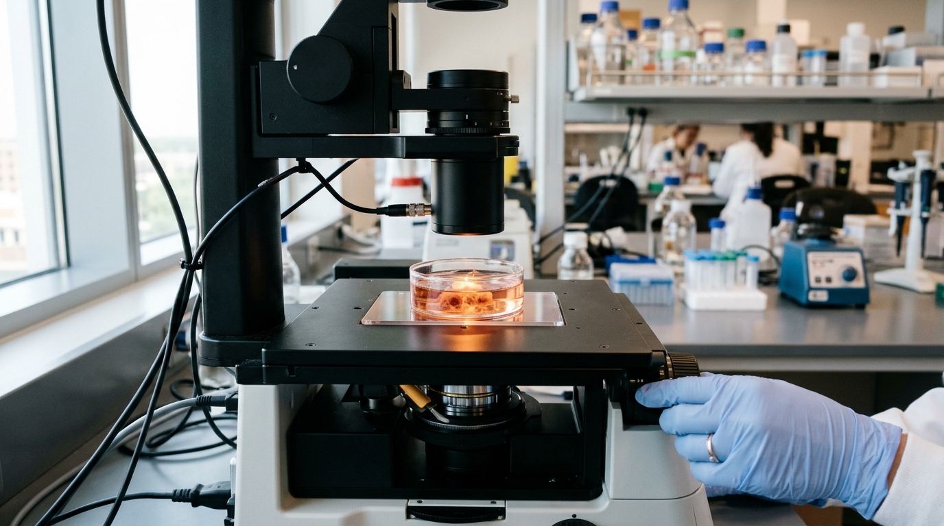
To trace how lipids move through a living tumor, the researchers injected fluorescently labeled low-density lipoprotein particles — the blood’s cholesterol carriers — into mice bearing pancreatic tumors. Using multiphoton intravital microscopy, they watched the labeled cholesterol in real time. Within the first hour, the lipid signal concentrated almost entirely in the cancer-associated fibroblasts rather than the cancer cells. By five hours, cholesterol had begun migrating from fibroblasts into carcinoma cells, accumulating precisely at the points where the two cell types touched. By roughly eighteen hours, the fibroblast signal had faded to near background while the cancer cells glowed strongly with the cholesterol label.
Flow cytometry of dissociated tumors confirmed this sequence: at ninety minutes after injection, PDGFRα-positive fibroblasts retained far more of the labeled LDL than cancer cells or myeloid cells, suggesting the fibroblasts were the first recipients of blood-derived lipid and then passed it along. When the team co-implanted fluorescently labeled human fibroblasts with mouse pancreatic cancer cells, the cancer cells acquired human fibroblast membrane markers within four days, and cholesterol fluorescence was twofold higher in cancer cells immediately adjacent to fibroblasts than in cancer cells farther away. The transfer, in other words, was contact-dependent — it collapsed almost completely when the cells were separated by a semipermeable membrane in transwell cultures, when the cultures were chilled to four degrees Celsius, or when calcium and magnesium were removed from the medium.
Imaging revealed the physical basis of the exchange. Fibroblasts and cancer cells formed sustained, synapse-like contacts, reminiscent of the immunological synapses that T cells form with their targets. At these junctions, fibroblast plasma membrane protrusions were visibly acquired by the touching cancer cells but not by bystander cells. Electron microscopy of gold-labeled fibroblasts and transfer of fluorescently tagged actin-containing protrusions confirmed that chunks of fibroblast membrane, not just soluble factors, crossed into the cancer cells. Notably, the dynamin inhibitor Dynasore did not block the uptake, arguing that the process is not conventional receptor-mediated endocytosis but true trogocytosis — the direct acquisition of membrane fragments from a neighboring cell.
The molecular trigger turned out to be phosphatidylserine exposure on the fibroblast surface. Blocking phosphatidylserine with recombinant annexin V or its mimic zinc-dipicolylamine significantly reduced membrane uptake, while manipulating the enzymes that control phosphatidylserine distribution shifted transfer in predictable directions: depleting the flippase scaffold TMEM30A, which increases surface phosphatidylserine, enhanced trogocytosis, whereas depleting the scramblase ANO6 nearly abolished it. Functionally, the consequences were dramatic. In lipid-depleted serum, cholesterol-auxotrophic pancreatic cancer cells underwent apoptosis, but coculture with patient-derived human fibroblasts rescued them — while fibroblast-conditioned medium or transwell coculture did not. Fibroblasts also sustained long-term clonogenic growth of cancer cells even when the LDL receptor was knocked down or the HDL receptor was blocked, indicating that this contact-based route operates independently of the classical lipoprotein uptake pathways.
The calcium signaling that activates ANO6 proved equally elegant. Only fibroblasts physically engaged with cancer cells showed calcium influx, detected with the sensors Fluo4 and GCaMP, and the signal vanished the moment the synapse-like contact broke. Pharmacological probing showed that store-operated calcium entry inhibitors BTP2 and zegocractin blocked the calcium rise, while a phospholipase C inhibitor did not, suggesting an ER store-independent activation of Orai channels. ER–PM junction markers, STIM1 and ANO6 itself, all polarized toward the heterotypic contact sites — a molecular architecture strikingly parallel to the immune synapse. In human data, high ANO6 expression was associated with worse overall survival in stage 1–3 pancreatic cancer patients in both the CPTAC protein and TCGA transcript datasets, and stromal ANO6 staining in 101 surgical samples correlated with poor tumor differentiation and inflammatory stromal signatures.
The therapeutic implications were tested with two repurposed drugs. Niclosamide and clofazimine, antimicrobial compounds originally identified as blockers of SARS-CoV-2-induced membrane fusion, both inhibit ANO6 scramblase activity. In mice bearing orthotopic pancreatic tumors, three weeks of oral treatment with either drug significantly reduced tumor weights. Niclosamide also suppressed the accumulation of injected BODIPY-cholesterol in cancer cells, whereas vehicle-treated tumors showed sixfold enrichment of the cholesterol label by sixteen hours. Genetically, cholesterol-auxotrophic tumor grafts grew significantly more slowly in Ano6-knockout hosts than in partially proficient mice. In the immune compartment, clofazimine-treated tumors contained fewer regulatory T cells, more CD8-positive T cells and more type I conventional dendritic cells, and the intratumoral cytotoxic T cells showed higher CD69, interferon-γ and granzyme B along with lower PD1 and TIM3 — a shift toward restored antitumor function.
The immunosuppressive arm of the mechanism was demonstrated directly in vitro. Cytotoxic T cells formed contacts with fibroblasts and trogocytosed their membranes, acquiring cholesterol in the process — a known driver of T cell exhaustion through lipotoxic stress. Blocking phosphatidylserine transfer with anti-TIM3 antibody or annexin V, or pretreating fibroblasts with clofazimine or niclosamide, reduced membrane uptake, lowered T cell cholesterol and restored interferon-γ production. Genetically depleting ANO6 in the fibroblasts reproduced all of these effects and increased the T cells’ ability to kill target cells. Tumor-infiltrating T cells in mice carried fibroblast membrane proteins on their surfaces, providing direct in vivo evidence that this transfer occurs inside tumors.
The authors are careful about the limits of their work. ANO6 is expressed by multiple cell types in the tumor, and the drugs have targets other than ANO6, so the pharmacological results alone cannot prove CAF-restricted target engagement — though the genetic experiments anchor the mechanism firmly to ANO6. Dependence on trogocytic lipid supply is also unlikely to be uniform across pancreatic cancers: basal-like cancer cell lines acquired fibroblast membrane more avidly than classical lines, and stromal ANO6 was lowest in tumors of the classical subtype. Pancreatic cancer cells retain other lipid acquisition routes, including macropinocytosis, receptor-mediated uptake and de novo lipogenesis, so trogocytosis should be viewed as one quantitatively important route among several rather than the sole supply line. Even so, the identification of a druggable, calcium-regulated switch that simultaneously feeds the tumor and disarms the immune system offers a compelling new entry point for treating desmoplastic, fibroblast-rich cancers — and the Orai inhibitor zegocractin, already in clinical trials for acute pancreatitis, could potentially be redeployed to disrupt these heterotypic cell contacts in patients.
Subject of Research: ANO6-dependent trogocytosis as a mechanism of lipid delivery and immune suppression in pancreatic cancer
Article Title: ANO6-mediated trogocytosis of cancer-associated fibroblasts promotes pancreatic cancer cell growth and immune suppression
Article References: Ogier, C., Charles Solomon, A. M., Bondarenko, E., Wu, Z., Lu, Z., Bui, Q. T., Dhamdhere, M. R., Tang, Y., Recoules, L., Klochkova, A., Gabitova-Cornell, L., Bayarmagnai, B., Restifo, D., Surumbayeva, A., Vendramini-Costa, D. B., Deneka, A. Y., Francescone, R., Lilly, A. C., Sipman, A., … Astsaturov, I. (2026). ANO6-mediated trogocytosis of cancer-associated fibroblasts promotes pancreatic cancer cell growth and immune suppression. Nature Cancer. https://doi.org/10.1038/s43018-026-01265-5
Image Credits: AI Generated
DOI: 10.1038/s43018-026-01265-5
Keywords: pancreatic cancer, cancer-associated fibroblasts, trogocytosis, ANO6, phosphatidylserine, cholesterol transfer, tumor microenvironment, immune suppression, cytotoxic T cells, niclosamide, clofazimine, calcium signaling
News Source: Nathaniel Bowman. (November 5, 2026). Fibroblasts Feed Pancreatic Tumors Through Contact-Based Lipid Transfer, Study Finds. Scienmag.