


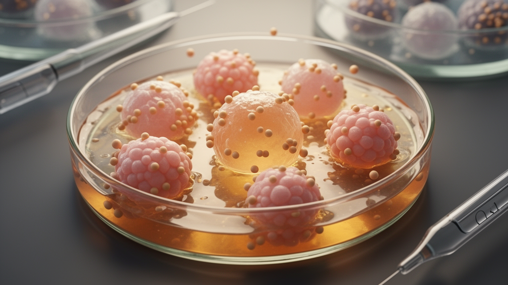
Patient-derived tumor organoids that retain immune cells are emerging as a promising preclinical platform for evaluating cancer immunotherapies, according to a review published in Frontiers in Oncology. The review, authored by researchers at Jilin Cancer Hospital in Changchun, China, examines four classes of tumor immune organoid models and compares their detection indicators, model fidelity, and experimental reproducibility. The authors argue that these three-dimensional culture systems address a persistent gap in precision tumor immunotherapy: the lack of functional in vitro models that can predict treatment efficacy while preserving the individual immune characteristics of a patient’s tumor.
Conventional preclinical tools fall short in specific ways. Two-dimensional cell lines lack three-dimensional architecture, cellular heterogeneity, and an intact tumor immune microenvironment, so they cannot accurately assess T cell infiltration, activation, and bystander killing mediated by multispecific antibodies. Patient-derived xenograft models, meanwhile, struggle to simultaneously restore the physiological states of the human immune system, tumor tissue, and the complex composition of the tumor microenvironment. The review states that deviations between results from these models and actual patient efficacy and toxicity responses contribute to the high failure rate of immunotherapy drugs in clinical trials. Organoids—self-organizing three-dimensional multicellular structures derived from stem and progenitor cells—retain key cellular components and partially reproduce the structural and functional features of the original tissue, and immune-functional versions add autologous immune populations and stromal components to recapitulate tumor-immune crosstalk.
The first model class is the air-liquid interface (ALI) organoid, pioneered across multiple cancer types by Neal and colleagues. Built on a double-layer collagen gel Transwell system, with the apical gel surface exposed to air and the basal side immersed in medium, ALI cultures preserve endogenous tumor-infiltrating immune cells and stromal fibroblasts, including T cells, B cells, NK cells, and macrophages. Critically, these organoids maintain the original T-cell receptor repertoire, clonal diversity, and expression profiles of tumor-infiltrating lymphocytes, and they replicate clinical checkpoint blockade: anti-PD-1/PD-L1 treatment expands tumor-specific T cells and induces cytotoxicity. In colorectal cancer, ALI organoids preserved native architecture, immune infiltration, and mutational landscapes, and nivolumab induced Treg expansion—a resistance hallmark absent in co-culture systems—suggesting that intact tumor immune interactions are essential for evaluating immunotherapy and dissecting resistance.
ALI models carry substantial limitations. The protocol is technically demanding, with low inter-laboratory reproducibility and no unified standardization. Long-term culture causes progressive viability loss and phenotypic drift in endogenous immune and stromal cells; persistent antigen stimulation, limited nutrient and oxygen diffusion, and the absence of systemic immune-cell replenishment may promote T cell dysfunction or exhaustion, marked by reduced cytotoxic and proliferative capacity and increased expression of inhibitory receptors such as PD-1, Tim3, and LAG3. The reliable window for immunotherapy testing is therefore likely concentrated in the early culture phase, though the precise timing depends on tumor type, donor characteristics, and experimental endpoints. Supplementation with cytokines such as IL-2, IL-7, or IL-15, oxygen-tension optimization, and extracellular-matrix adjustments may mitigate immune-cell decline, but the review notes these interventions require further experimental validation. The model also lacks vasculature, fluid shear stress, and systemic immune circulation, and its format is incompatible with high-throughput screening.
Microfluidic organoid-on-a-chip platforms offer a different solution. In traditional Matrigel embedding, nutrient supply and waste removal rely on passive diffusion, which becomes inadequate as organoids grow, leaving central regions starved of oxygen. Continuous perfusion replenishes oxygen and nutrients while removing metabolic products, creating more stable growth conditions and improving suitability for high-throughput screening. The review highlights several platforms: the chip-free droplet microfluidic system OsciSphere, which achieves uniform bioassembly of three-dimensional multicellular systems and supports quantitative evaluation of PD-1 blockade immunotherapy; microfluidic devices used to test an oncolytic adenovirus in patient-derived pharyngeal and breast cancer organoids; and a platform for scarce pancreatic cancer biopsy samples that enables chemotherapy screening and NK cell-based combination immunotherapy evaluation. However, flow-induced shear stress may compromise immune-cell survival or alter immune-cell phenotype, with effects depending on cell subtype, exposure duration, and flow field, so flow conditions must be optimized through computational modeling and experimental calibration. The authors also flag practical barriers: microfluidic systems handle viscous matrices like Matrigel poorly, dynamic perfusion demands specialized expertise, and customized equipment costs can be prohibitive for many laboratories.
A second major category reconstructs immunity by adding cells rather than preserving them. Tumor organoid–PBMC co-culture systems pair patient-derived organoids with peripheral blood mononuclear cells in several formats: fully embedded Matrigel cultures that permit T cell and NK cell infiltration, partitioned systems that isolate organoids from PBMCs to study cytokine-mediated indirect responses, and direct co-cultures without Matrigel that rapidly generate tumor-specific effector T cells. In one standardized two-stage workflow, tumor-reactive T cells from peripheral blood are expanded over an initial two-week co-culture with interferon-γ-stimulated autologous tumor cells, then functionally evaluated for cytokine secretion, degranulation, and killing of tumor organoids. In non-small cell lung cancer and microsatellite-unstable colorectal cancer samples, the system cultivated tumor-responsive CD8+ T cell populations within two weeks, with a success rate of approximately 33 to 50 percent—a figure influenced by biopsy quality, disease stage, initial cell yield, tumor mutational burden, and the presence of pre-existing tumor-reactive T cell clones in peripheral blood.
Organoid–TIL co-culture systems take a more tumor-specific approach, isolating CD45+ immune cells from digested surgical specimens and co-culturing purified autologous tumor-infiltrating lymphocytes with matched organoids. Applications reported in the review include a colorectal cancer system that revealed time-dependent, actin- and PI3K-dependent transfer of lymphocyte membrane proteins to tumor cells; a microsatellite instability-high colorectal cancer model linking post-treatment local inflammation and neutrophil-induced immunosuppression to blunted checkpoint blockade efficacy and higher progression risk; an ex vivo platform that rapidly identifies tumor-specific T cell receptors to support TCR-T immunotherapy development; and an individualized platform pairing expanded autologous TILs with matched colorectal cancer organoids to predict clinical benefit of neoadjuvant chemoradiotherapy. The review cautions, however, that PBMCs differ from TILs in T cell receptor diversity and phenotype, that tumor-specific clones are typically rarer in peripheral blood than at the tumor site, and that circulating suppressive populations such as myeloid-derived suppressor cells and Tregs can skew co-culture responses.
More elaborate architectures attempt to rebuild the full tumor microenvironment. Bottom-up reconstruction assembles tumor organoids, cancer-associated fibroblasts, endothelial cells, and macrophages—derived from autologous PBMCs or sorted from tumor tissue—in defined ratios within three-dimensional scaffolds, microfluidic chips, or ultra-low adhesion plates. Reported findings include a multicellular pancreatic cancer co-culture showing that CSF1R inhibitor-mediated elimination of M2-polarized macrophages reverses microenvironment-mediated chemotherapy resistance, and a gravity-perfused immune-infiltrating tumor spheroid model that induced time-dependent, tissue-specific monocyte recruitment in breast and liver tumor spheroids within a week. Three-dimensional bioprinting adds precise spatial control, reproducing tumor heterogeneity, nutrient gradients, hypoxic regions, and multicellular spheroids with necrotic cores. But these systems face their own constraints: macrophages drift phenotypically in short-term culture, cell ratios are difficult to standardize, tumor cells and stromal cells have conflicting growth-factor requirements, vascular networks show incomplete lumen formation and limited pericyte coverage, and bioprinting shear stress can damage cells or trigger unintended immune activation.
The review is explicit that clinical translation remains premature. Most current immune organoid systems support only short-term co-culture of one to two weeks, insufficient for modeling durable immune memory, exhaustion reversal, or chronic resistance; selected studies using perfusion bioreactors and cytokine supplementation have extended immune-cell viability to roughly three to four weeks, but prolonged cytokine exposure may itself promote terminal differentiation and exhaustion-marker expression. Prospective clinical validation of organoid-based immunotherapy prediction remains limited: most studies have involved small, single-center cohorts without validated clinical prediction models, and standardized protocols with predefined clinical endpoints are lacking. Severe cytokine release syndrome—a multi-organ systemic inflammatory response—cannot be fully recapitulated in isolated organoid cultures, so the authors position these platforms as complementary to, rather than replacements for, systemic toxicity evaluation. Looking forward, they propose artificial intelligence-assisted image analysis, single-cell and spatial multi-omics integration, CRISPR gene engineering, and microenvironment engineering incorporating lymphatic endothelial cells, nerve fibers, and vascular networks as routes to improve fidelity, standardization, and scalability, with larger multicenter prospective studies required to determine whether organoid assays can reliably predict durable patient benefit.
Subject of Research: Tumor immune organoids as preclinical models for evaluating cancer immunotherapy efficacy
Article Title: Tumor immune organoids support preclinical evaluation of cancer immunotherapies through comparative modelling and translational investigation
Article References: Zhang, Y., Wang, Z., Xiu, Y., Wang, H., Zhao, K., & Chen, Z. (2026). Tumor immune organoids support preclinical evaluation of cancer immunotherapies through comparative modelling and translational investigation. Frontiers in Oncology, 16, Article 1988538. https://doi.org/10.3389/fonc.2026.1988538
Image Credits: AI Generated
DOI: 10.3389/fonc.2026.1988538
Keywords: tumor organoids, immunotherapy, tumor microenvironment, immune checkpoint inhibitors, patient-derived models, organoid-on-a-chip, TIL co-culture, 3D bioprinting, preclinical models, precision oncology, PBMC co-culture, drug resistance
News Source: Nathaniel Bowman. (October 29, 2026). Lab-Grown Tumor Organoids With Immune Cells Aim to Predict Immunotherapy Response. Scienmag.



