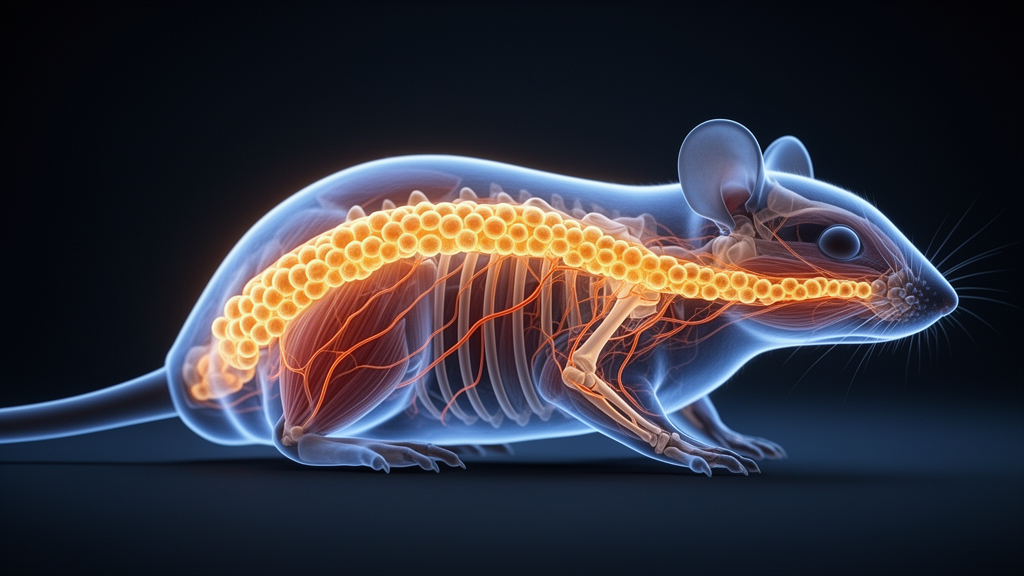
Researchers have built a genetically engineered mouse line that switches on a fluorescent marker almost exclusively in the small blood vessels of muscle and fat, giving vascular biologists a precision instrument they have long lacked.
Endothelial cells form the thin inner lining of blood and lymphatic vessels and do far more than act as a passive barrier. They help regulate vascular tone, inflammatory responses, clot formation and the growth of new vessels, and endothelial dysfunction is a hallmark of systemic conditions including atherosclerosis, diabetes and heart failure. Crucially, endothelial cells are not all alike. Their properties differ markedly between vascular beds and tissues, and this heterogeneity is especially pronounced in skeletal and cardiac muscle, where the vasculature must meet high metabolic demands, adjust blood flow during contraction and support repair after injury. In muscle, vascular endothelial cells form a functional unit with muscle fibers and satellite cells that contributes to metabolic homeostasis, regenerative capacity and adaptation to exercise.
Studying these specialized cells has been hampered by a shortage of genetic tools. Cre recombinase mouse lines, particularly inducible ones, allow precise spatial and temporal control of gene expression, but the widely used lines driven by the Tie2 or VE-Cadherin (Cdh5) promoters target endothelial cells broadly across many organs and lack tissue specificity. Tissue-specific endothelial Cre lines remain scarce; one of the few examples is Slco1a4-CreERT2, which targets the cerebrovascular endothelium. Until now, no Cre line existed that specifically targeted the endothelial cells of muscle.
The team behind the new line began with data rather than animal breeding. By mining publicly available single-cell RNA sequencing datasets, they searched for genes that might distinguish muscle endothelial cells from other cell types. Analysis of the Murine Endothelial Cell Atlas, which profiles endothelial cells from 11 adult mouse tissues, revealed abundant CTRP9 expression in endothelial cells from the heart and limb skeletal muscle, with minimal expression in non-muscular organs. Within the heart, CTRP9 was concentrated in microvascular endothelial cells, including capillary, capillary arteriolar and capillary venular populations, and was reduced in arterial, large-vessel venous and lymphatic endothelial cells. In skeletal muscle, the gene was broadly expressed across capillary endothelial subtypes but lower in large-artery endothelial cells. A parallel analysis showed similar enrichment of CTRP9 in the vascular endothelium of adipose tissue, with minimal expression in other fat-resident cell types.
To check that this signal really belonged to blood vessels rather than the surrounding tissue, the researchers turned to the Tabula Muris dataset, a single-cell transcriptomic atlas covering 20 mouse organs and 38 cell types. In both heart and limb skeletal muscle, CTRP9 expression was confined to endothelial cells, with little to none in cardiomyocytes, fibroblasts, smooth muscle cells or immune cells. This finding also corrects an earlier picture of the gene’s biology. CTRP9, short for C1q/tumor necrosis factor-related protein-9, is an adiponectin paralog that acts as an adipokine; after proteolytic cleavage it releases a globular domain that circulates in the blood and, through adiponectin receptor 1 and N-cadherin, triggers signaling pathways that modulate glucose and lipid metabolism, vasodilation and cell differentiation. An earlier study concluded that CTRP9 expression in mice was restricted to adipose tissue, but that work relied on bulk tissue qPCR, which measures whole-tissue homogenates and cannot resolve which cells produce the protein. The single-cell and reporter data now indicate that, although CTRP9 was initially recognized as an adipokine, it is primarily produced by microvascular endothelial cells in muscle and adipose tissues, a distribution that aligns with its reported effects on skeletal muscle cells, cardiac cells, adipocytes and endothelial cells.
With the marker identified, the team built the tool. Using CRISPR/Cas9-mediated genome editing, they inserted a CreERT2-P2A-tdTomato-WPRE-polyA cassette immediately downstream of the translation start codon in exon 2 of the CTRP9 locus, flanked by 1,645-base-pair 5′ and 1,595-base-pair 3′ homology arms. The P2A self-cleaving peptide allows CreERT2 and the red fluorescent tdTomato reporter to be expressed independently, so tdTomato mirrors endogenous CTRP9 expression. The donor vector, guide RNA and Cas9 mRNA were co-microinjected into fertilized mouse oocytes on a C57BL/6J background. PCR analysis confirmed the expected 2,207-base-pair and 1,991-base-pair bands in heterozygous knock-in animals but not in wild-type controls, and Southern blot analysis verified precise integration, yielding a 14.87-kilobase fragment with EcoNI digestion and a 7.58-kilobase fragment with BstEII digestion. Sequencing of 16 predicted off-target sites found no detectable mutations or genomic alterations.
The tdTomato reporter alone already mapped CTRP9 expression across development. At embryonic day 14.5, tdTomato appeared in a few cells in the upper cervical spinal cord and heart, where it co-localized with the endothelial marker CD31, showing CTRP9 activity in the embryonic vasculature. At postnatal day 12 and day 60, tdTomato-positive cells were found in the heart, limb skeletal muscle and tongue, a specialized muscular organ, and the majority or all of them co-expressed CD31, confirming that CTRP9 is expressed mainly in the vascular endothelium of muscular tissues from the postnatal period into adulthood.
To test the inducible Cre system itself, the knock-in mice were crossed with Rosa26-LSL-EYFP reporter mice. In these double-transgenic animals, the CreERT2 fusion protein carries a mutated estrogen receptor domain that ignores endogenous estrogens but is activated by tamoxifen; without the drug, CreERT2 stays sequestered in the cytoplasm, and once tamoxifen is given it moves into the nucleus, removes the loxP-flanked STOP cassette and permanently switches on EYFP from the Rosa26 locus. Tamoxifen was given at postnatal day 7 for five consecutive days with analysis at day 12, or for seven consecutive days starting at seven weeks of age with analysis at day 60. Mice that received no tamoxifen showed no EYFP signal in the heart, skeletal muscle, tongue or adipose tissue, demonstrating the absence of tamoxifen-independent recombination. RNA in situ hybridization further confirmed that all CTRP9-positive cells showed EYFP expression, meaning the reporter faithfully reflects endogenous gene activity.
Quantitative immunofluorescence then defined exactly which endothelial subsets the line captures. In the heart, all EYFP-positive cells expressed CD31, and EYFP-positive endothelial cells made up 63.60 percent plus or minus 2.84 percent of CD31-positive cells at day 12 and 60.56 percent plus or minus 1.14 percent at day 60. Using alpha-smooth muscle actin and EphB4 to mark arterial and venous endothelial cells respectively, the labeled population overlapped only partially with these compartments: EYFP-positive arterial cells accounted for 19.98 percent plus or minus 3.72 percent of arterial endothelial cells at day 12 and 14.12 percent plus or minus 0.65 percent at day 60, while EYFP-positive venous cells accounted for 33.68 percent plus or minus 2.48 percent and 31.64 percent plus or minus 2.56 percent of venous endothelial cells. The pattern points to a preference for microvascular endothelial cells. In skeletal muscle, labeled cells represented 79.21 percent plus or minus 2.89 percent of endothelial cells at day 12 and 62.92 percent plus or minus 2.38 percent at day 60; in the tongue, 63.08 percent plus or minus 2.31 percent and 77.90 percent plus or minus 2.05 percent. In adipose tissue, EYFP-positive endothelial cells reached 76.96 percent plus or minus 2.58 percent at day 12 and 80.90 percent plus or minus 1.20 percent at day 60. By contrast, the stomach and small intestine, both rich in smooth muscle, showed minimal endothelial labeling, and the testis and brain showed no detectable EYFP or tdTomato signal at all, underscoring the restriction of the line to muscle and adipose tissues.
Finally, the researchers asked whether the labeling holds up under the stress of tissue repair. They induced muscle injury by injecting barium chloride into the tibialis anterior muscle of tamoxifen-treated mice and examined the tissue five days later. EYFP-positive cell numbers rose in the injured muscle, every labeled cell expressed CD31, and every proliferating endothelial cell, marked by CD31 and Ki67 co-expression, was EYFP-positive, indicating that the newly formed endothelial cells of regenerating muscle originated from the CTRP9-positive microvascular population. The labeled cells never expressed F4/80, a macrophage marker, so the line stayed strictly endothelial-specific even under injury conditions. The authors position the CTRP9-CreERT2 mouse as a resource for precisely manipulating this endothelial subset, enabling more targeted investigations of endothelial diversity and vascular function in muscle metabolism, regeneration and disease. As with any new genetic tool, its findings rest on mouse models and reporter-based measurements, and its usefulness in specific experimental contexts will be tested by the laboratories that adopt it.
Subject of Research: Generation and characterization of a CTRP9-CreERT2 knock-in mouse line targeting microvascular endothelial cells in muscle and adipose tissues
Article Title: The generation and characterization of CTRP9-CreERT2 knock-in mice for targeting endothelial cell muscle and adipose tissues
Article References: Wang, Q., Fu, Z., Wang, H., Ao, H., Li, S., Xu, J., Meng, Y., Duan, Q., Yang, X., Wang, J., & Teng, Y. (2026). The generation and characterization of CTRP9-CreERT2 knock-in mice for targeting endothelial cell muscle and adipose tissues. Cell Regeneration, 15(1), Article 29. https://doi.org/10.1186/s13619-026-00300-1
Image Credits: AI Generated
DOI: 10.1186/s13619-026-00300-1
Keywords: CTRP9, CreERT2, knock-in mice, endothelial cells, microvasculature, CRISPR/Cas9, single-cell RNA sequencing, skeletal muscle, adipose tissue, muscle regeneration, tdTomato reporter, vascular biology
News Source: Juliet Wilcox. (October 28, 2026). New mouse tool targets microvascular endothelial cells in muscle and fat. Scienmag.