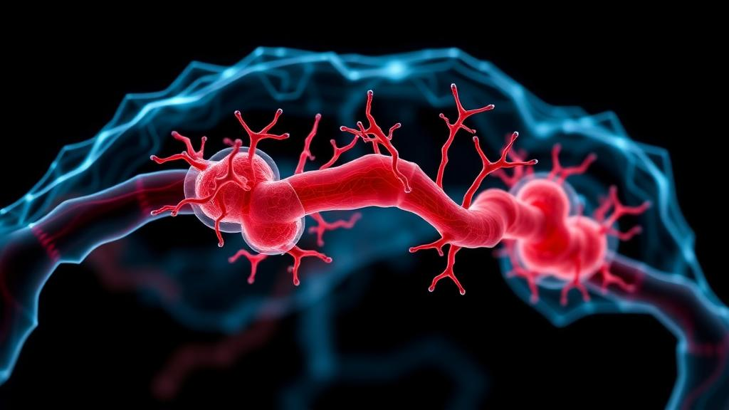
Moyamoya disease is one of the most enigmatic disorders in neurology. The distal segments of the internal carotid arteries, the main pipelines feeding the brain, slowly narrow and eventually occlude, while the brain compensates by sprouting a fragile, smoke-like network of collateral vessels at the base of the skull. The Japanese surgeons who first described the condition in 1969 named it moyamoya, meaning puff of smoke, after the hazy tangle of vessels that appears on angiograms. Despite decades of study, the mechanisms driving this progressive stenosis and the formation of these unstable collaterals remain poorly understood, in large part because the disease has been extraordinarily difficult to model in the laboratory.
Now a review published in Molecular Biology Reports by a team of Korean researchers led by Jong-Tae Kim, Dong Hyuk Youn, Jae Jun Lee and Jin Pyeong Jeon of Hallym University College of Medicine maps out how patient-derived induced pluripotent stem cells and organoids are transforming the field. The review traces the evolution of Moyamoya modeling from early studies of circulating endothelial progenitor cells to sophisticated three-dimensional vascular and neurovascular organoids, and argues that these platforms could finally capture the genetic, vascular and neurovascular complexity that conventional models have missed. The work arrives at a moment when the field’s central susceptibility gene, RNF213, is being dissected at molecular resolution, and when organoid technology is mature enough to reproduce vessel-like structures in a dish.
The genetic foundation of the disease was laid in 2011, when genome-wide association studies identified RNF213, which encodes the ring finger protein 213, as the first Moyamoya disease gene. The R4810K variant, a single amino acid substitution in East Asian patients, remains the strongest susceptibility factor, and subsequent genome-wide studies have uncovered additional loci. Yet the genotype-phenotype relationship is far from simple. Not every carrier develops disease, penetrance is incomplete, and European patients often present with Moyamoya angiopathy without the East Asian founder mutation. This complexity has made it clear that RNF213-dependent and RNF213-independent mechanisms both contribute, and that understanding them requires models built on human cells carrying the actual patient genome.
Induced pluripotent stem cells, or iPSCs, offer exactly that opportunity. By reprogramming peripheral blood mononuclear cells or skin fibroblasts from patients back into a pluripotent state, researchers can generate an unlimited supply of genetically matched cells and then differentiate them into any vascular lineage of interest. The first Moyamoya-specific iPSC lines were established in 2016 from patients with ischemic and hemorrhagic forms of the disease, followed in 2021 by a line carrying a confirmed RNF213 mutation. Validation of these lines requires confirming pluripotency markers, normal karyotype, and retention of the disease genotype, and once established, they can be directed toward endothelial cells, vascular smooth muscle cells, and related vascular lineages that constitute the arterial wall.
Two-dimensional iPSC-based models have already yielded concrete mechanistic insights. In 2013, researchers showed that the RNF213 R4810K variant downregulates securin, reducing the angiogenic activity of iPSC-derived vascular endothelial cells from Moyamoya patients, and that the same variant induces genomic instability through mitotic abnormalities. Later work demonstrated that RNF213 regulates endothelial barrier function, that its loss of function promotes pathological angiogenesis through the Hippo pathway, and that RNF213 knockdown combined with inflammation drives angiogenesis and phenotype switching via the JAK2/STAT3 pathway. Studies of iPSC-derived vascular smooth muscle cells revealed abnormal behavior compared with healthy controls, and comparative transcriptomic analysis of endothelial and smooth muscle lineages highlighted disrupted vascular cell interactions. Additional findings point to autophagic impairment, mitochondrial dysfunction, and altered responses to vascular endothelial growth factor and transforming growth factor-beta as contributing factors.
These 2D models, however, have inherent limitations. They flatten the vascular wall into a single layer, remove the mechanical forces of blood flow, and eliminate the cellular heterogeneity and spatial organization that define living vessels. Histopathological studies of Moyamoya arteries show intimal thickening, proliferation and migration of smooth muscle cells, and infiltration of macrophages and T cells, features that a flat culture cannot reproduce. The review therefore highlights the emerging use of three-dimensional vascular organoids, which self-organize into vessel-like networks embedded in an extracellular matrix, and neurovascular organoids, which integrate brain cells with vascular components to model the neurovascular unit, the interface where endothelial cells, pericytes, astrocytes and neurons meet at the blood-brain barrier.
Recent organoid studies are beginning to capture key features of Moyamoya vasculopathy. In 2026, researchers reported that RNF213 deficiency in human iPSC-derived vascular organoids reproduces a central feature of the disease’s vasculopathy, and a separate organoid modeling study combined with single-cell RNA sequencing revealed pathological smooth muscle cell migration, a hallmark of Moyamoya arteriopathy. Vascularized brain organoids, engineered through strategies that co-culture endothelial cells with neural progenitors or embed vascular lineages within brain organoid matrices, now allow researchers to study how Moyamoya-relevant vascular defects interact with brain tissue. These models reproduce cellular heterogeneity, vessel-like organization, and neurovascular unit interactions far more closely than conventional 2D cultures, providing a more integrated platform for investigating the complex vascular pathophysiology of the disease.
The review also points toward future directions that could sharpen these models further. Integration of multi-omics profiling, including single-cell RNA sequencing, proteomics and metabolomics, with patient-derived iPSC and organoid platforms could clarify genotype-phenotype relationships and identify disease-relevant molecular pathways. CRISPR-mediated genome editing offers the ability to introduce or correct RNF213 variants in isogenic cell lines, allowing researchers to isolate the effect of specific mutations against an otherwise identical genetic background. Organ-on-a-chip systems that apply shear stress and pulsatile flow could add the biomechanical dimension that static cultures lack, and adeno-associated viral vectors and lipid nanoparticles could enable therapeutic gene delivery studies within organoid models. Discordant monozygotic twin studies, which have already profiled circulating microRNAs and endothelial microRNA expression using iPSC lines, add another layer of control by isolating genetic from environmental contributions.
With further standardization and validation, iPSC- and organoid-based models could support mechanistic studies, therapeutic screening, and personalized disease modeling, in which a patient’s own cells are used to test drug responses before treatment begins. The review underscores that these technologies are not merely incremental improvements but represent a fundamental shift in how a rare cerebrovascular disorder can be studied, moving from indirect proxies of vascular dysfunction to direct, human, genetically faithful models of the disease itself. For a condition that has resisted mechanistic dissection for more than half a century, the convergence of patient-derived stem cells, organoids, genome editing and multi-omics profiling may finally provide the tools needed to unravel why cerebral arteries narrow, why fragile collaterals form, and how both processes might one day be prevented.
Subject of Research: Moyamoya disease modeling with patient-derived iPSCs and organoids
Article Title: Modeling Moyamoya disease using patient-derived induced pluripotent stem cells and organoids
Article References: Kim, J.-T., Youn, D. H., Jung, H., Lee, J. H., Hong, E. P., Kim, Y., Cho, K.-W., Lee, J. J., & Jeon, J. P. (2026). Modeling Moyamoya disease using patient-derived induced pluripotent stem cells and organoids. Molecular Biology Reports, 53(1), Article 1750. https://doi.org/10.1007/s11033-026-12932-2
Image Credits: AI Generated
DOI: 10.1007/s11033-026-12932-2
Keywords: Moyamoya disease, induced pluripotent stem cells, organoids, RNF213, disease modeling, vascular organoids, brain organoids, endothelial cells, vascular smooth muscle cells, CRISPR genome editing, neurovascular unit, single-cell RNA sequencing
News Source: Juliet Wilcox. (October 18, 2026). Patient-derived stem cells and organoids open a new window on Moyamoya disease. Scienmag.