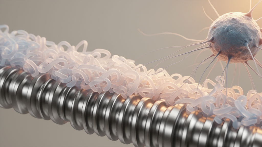
Every bone implant undergoes a transformation within seconds of entering the body. Long before osteoblasts arrive or new mineral begins to form, the surface of a titanium screw, a ceramic scaffold, or a polymer graft is coated by a layer of proteins drawn from blood, marrow fluid, and surrounding tissue. A new critical review published in Frontiers in Bioengineering and Biotechnology argues that this acquired protein layer, rather than the material itself, may be the true biological identity of a bone-regenerative implant, and that the field has been measuring the wrong things for decades.
The review, led by Yunhe Zhang of Dalian Medical University with colleagues from China Medical University and Haicheng Orthopedics Hospital, examines how the quantity, identity, orientation, conformation, and exchange kinetics of adsorbed proteins collectively determine what cells actually see when they encounter an implant. The authors introduce the term interfacial protein state to describe this measurable combination, and they propose that researchers report all five dimensions together under matched conditions rather than relying on total protein mass alone. Their central argument is that two surfaces carrying identical amounts of fibronectin can produce entirely different biological outcomes depending on whether the integrin-binding RGD motif and the PHSRN synergy site remain accessible to cellular receptors.
The evidence for this principle is strongest in defined laboratory systems. In one frequently cited comparison, fibronectin adsorbed onto poly(ethyl acrylate) and poly(methyl acrylate) surfaces reached similar surface densities of approximately 340 nanograms per square centimeter when exposed to a 20 microgram per milliliter coating solution for ten minutes. Yet atomic force microscopy revealed fibrillar assembly on one polymer and a more globular organization on the other, and an antibody-based accessibility assay showed that the fraction of fibronectin molecules presenting their integrin-binding domain differed between the two surfaces. The protein was there in equal amounts on both materials, but it was not equally available to the cells that needed to read it.
This distinction between quantity and functional presentation has direct consequences for how bone implants are designed and evaluated. The review emphasizes that increasing the amount of an adsorbed protein does not restore signaling if the critical domain is turned toward the material or sterically blocked by neighboring molecules. Orientation, the authors argue, can change function even when protein identity and abundance remain unchanged. Receptor-binding regions that face the surrounding fluid remain available to cells, while the same regions become biologically silent when they lie against the material surface. This principle applies not only to fibronectin and vitronectin but also to the Fc region of immunoglobulins and to receptor-recognition sites on complement fragments.
Conformation presents an equally complex picture. The review distinguishes between limited structural extension that can preserve or even enhance physiological function and deeper denaturation that exposes nonphysiological epitopes and triggers inflammatory pathways. Fibronectin illustrates the potentially beneficial side: after adsorption onto certain surfaces, it can transition from a compact form toward an extended or fibrillar organization that makes integrin-binding and growth-factor-binding regions more accessible. Fibrinogen demonstrates the opposite direction. When adsorption unfolds this coagulation protein excessively, the gamma-chain region recognized by the Mac-1 integrin on leukocytes becomes exposed, driving inflammatory-cell adhesion and cytokine release. Albumin, often assumed to be a passive blocking agent, can acquire affinity for scavenger receptors when sufficiently denatured.
The temporal dimension adds another layer of complexity. The review documents that the protein layer is not frozen after implantation but continuously evolves through competitive exchange, enzymatic cleavage, cell-mediated remodeling, and deposition of new matrix. In nanoparticle systems, a complex plasma protein corona has been detected within less than thirty seconds of exposure. Human serum albumin dissociation from a specific particle type showed a half-life of approximately thirty minutes, though the authors stress this is a protein-and-material-specific result rather than a universal timescale. By the time immune cells, endothelial cells, and osteoprogenitors reach the implant surface, they are contacting a material-protein composite that has already undergone multiple rounds of exchange and restructuring.
One of the most clinically relevant sections addresses autologous blood-product precoatings. Platelet-rich plasma and platelet-rich fibrin are widely used to deliver patient-derived proteins and growth factors to implant surfaces before insertion. The review acknowledges that these preparations differ substantially in their growth-factor release profiles depending on centrifugation protocol, anticoagulation, activation state, and fibrin organization. However, it also notes that a randomized split-mouth trial involving fifteen patients and thirty dental implants found no statistically significant between-group differences in assessed outcomes after one year when liquid platelet-rich fibrin was applied to implant surfaces. The authors interpret this as limiting unqualified efficacy claims rather than excluding benefits under other protocols, and they emphasize that neither growth-factor content nor clinical application alone validates a particular protein-state mechanism.
The review also confronts an uncomfortable reality about bacterial competition. Staphylococcal fibronectin-binding proteins can exploit adsorbed fibronectin to engage host alpha-5 beta-1 integrins, while fibrinogen-binding adhesins support bacterial attachment to conditioned surfaces. The very protein features that support mammalian-cell attachment may simultaneously facilitate bacterial colonization and biofilm development. The authors argue that host-cell adhesion alone cannot establish resistance to bacterial attachment and that protein-conditioned materials require parallel assessment with clinically relevant bacterial species. They cite an experimental system that combined fibronectin and BMP-2 presentation on coated titanium nanotopography with examination of both mesenchymal stromal cells and Pseudomonas aeruginosa in the same material platform, demonstrating that host-cell support and biofilm inhibition can be evaluated together.
Perhaps the most sobering section of the review addresses what remains unvalidated. The authors are explicit that predicting bone-healing outcomes across different materials, guiding patient-specific implant selection, and replacing growth-factor delivery through endogenous protein capture all remain unproven hypotheses. They distinguish carefully between evidence obtained with purified protein coatings, recombinant growth factors, and dispersed nanoparticles versus evidence of endogenous protein recruitment at a bone-contacting implant. A mechanism supported only by in vitro systems, non-bone animal models, or nanoparticle studies is not described as clinically established without cross-scale validation. The review proposes a six-level evidence ladder ranging from interface formation through receptor translation to human translation, with each level requiring progressively stronger causal evidence.
The authors conclude that the interfacial protein state framework is a reporting and comparison scheme rather than a validated predictor or clinical score. They call for future studies to connect measurements on the final sterilized product to causal perturbations, disease-relevant biofluids, safety endpoints, and indication-matched outcomes. Patient-specific implant selection and programmed endogenous protein exchange remain research proposals until prospective validation demonstrates added clinical value beyond conventional variables. What the review does establish is that the protein layer acquired by a bone implant within minutes of insertion is not a passive footnote to material design but an active biological interface that determines whether the body builds bone, mounts inflammation, or invites bacterial colonization at the site of repair.
Subject of Research: How endogenous protein adsorption and functional presentation at bone-regenerative biointerfaces control receptor recognition, inflammation, and bone healing outcomes
Article Title: Endogenous protein adsorption and functional presentation at bone-regenerative biointerfaces
Article References: Zhang, Y., Xu, Y., Zhang, C., & Lu, B. (2026). Endogenous protein adsorption and functional presentation at bone-regenerative biointerfaces. Frontiers in Bioengineering and Biotechnology, 14, Article 1965344. https://doi.org/10.3389/fbioe.2026.1965344
Image Credits: AI Generated
DOI: 10.3389/fbioe.2026.1965344
Keywords: protein corona, bone regeneration, biointerface, fibronectin, fibrinogen, integrin signaling, BMP-2, titanium implants, osteogenesis, protein adsorption, biomaterials, osteoimmunology
News Source: Denise Maddox. (October 16, 2026). The Hidden Protein Layer That Decides Whether a Bone Implant Heals or Fails. Scienmag.