Colorectal cancer prognosis has long been judged mainly by tumor stage and spread, but the immune cells living inside and around the tumor may carry additional information.
The work, led by Yan Lai, Juan Wang and colleagues with Cheng Yang as a senior author, focused on a persistent gap in cancer immunology. Scientists know that the tumor microenvironment influences how colorectal cancer progresses, and that spatial immune aggregates, or SIAs, are part of that environment. What has remained unclear is the prognostic relevance of spatially resolved immune subsets, particularly phenotypes involving B cells, and how those features compare against established clinicopathological predictors when both are tested rigorously in the same cohort.
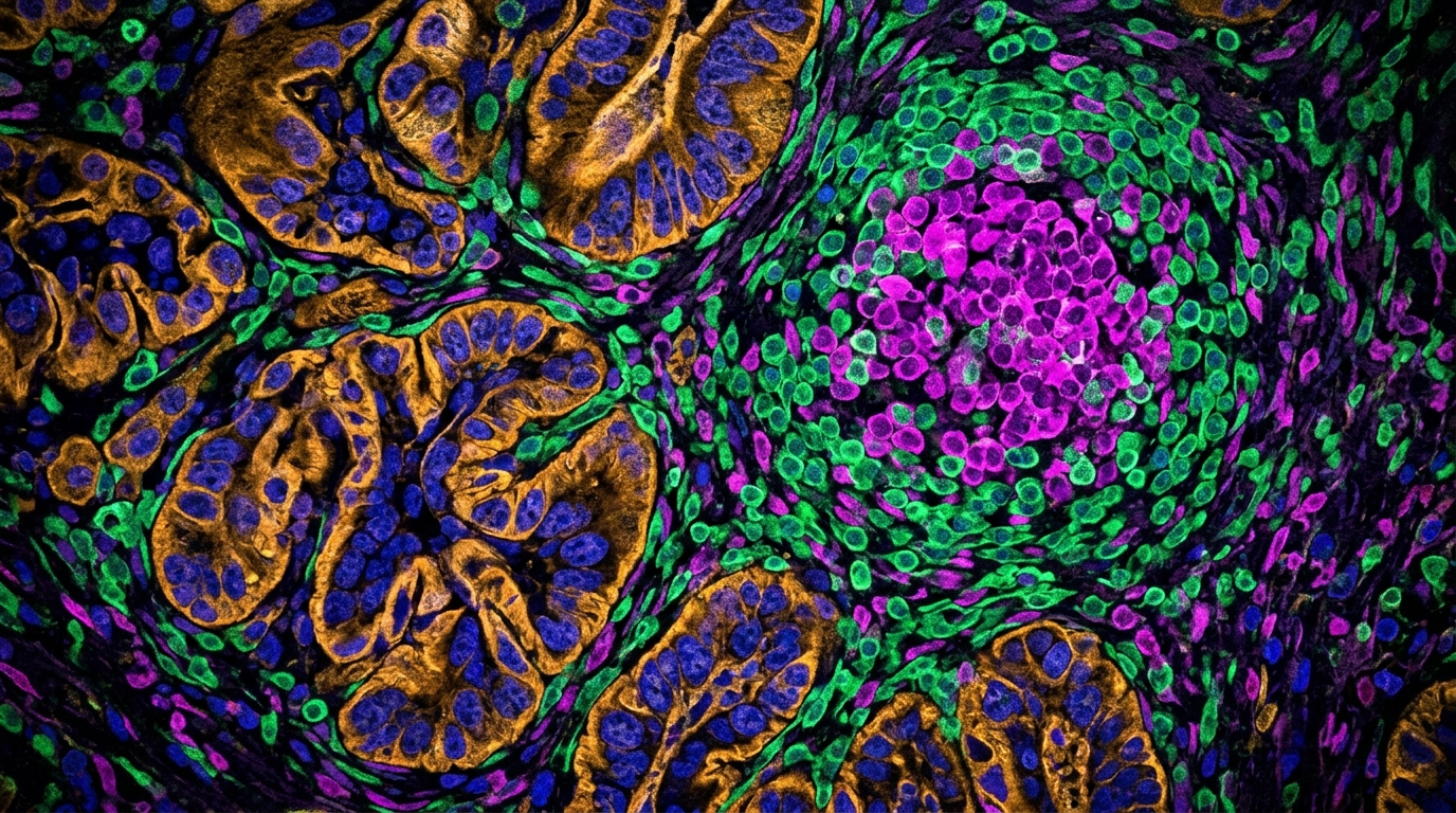
Technically, the pipeline was elaborate. Tissue sections were stained with a panel of antibodies marking ten immune phenotypes, and a deep learning algorithm segmented every cell individually. A biologically informed version of the density-based spatial clustering algorithm DBSCAN then detected spatial immune aggregates automatically, without relying on manual annotation. The team validated this automated detection against annotations reviewed by pathologists and reported high concordance, an important step because any downstream spatial analysis depends on the accuracy of the underlying cell map.
Among the ten phenotypes, one cell type dominated the tumor microenvironment: non-proliferating CD3-positive, CD8-negative, FOXP3-negative T cells made up 64.9 percent of the immune landscape. Immune composition was not static across the cohort. It varied with disease stage and with metastatic status, suggesting that the immune ecosystem shifts as colorectal cancer advances, though the study characterized these associations rather than establishing causal mechanisms.
The spatial analysis produced several region-specific findings. Within spatial immune aggregate regions, proliferating CD8-positive T cells were associated with favorable patient outcome, a pattern consistent with the idea that actively dividing cytotoxic T cells concentrated near organized immune structures accompany better disease course. In contrast, non-proliferating CD21-positive B cells located outside spatial immune aggregate regions were associated with poorer outcome. This B-cell finding is notable because B-cell-related phenotypes have been incompletely defined in colorectal cancer prognosis research, and the result points to a specific spatial context, non-SIA regions, where these cells carry adverse prognostic weight.
Spatial proximity analysis added another layer. The researchers identified distance patterns associated with metastatic status, including shortened distances between CD21-negative B cells and the dominant non-proliferating CD3-positive, CD8-negative, FOXP3-negative T cell population in patients with metastatic disease. Such proximity measurements are only possible with single-cell spatial resolution, which is precisely what the multiplex immunohistochemistry and segmentation pipeline provided.
The most sobering part of the study came from its prognostic benchmarking. The authors restricted survival modeling to the 636 patients with complete survival information and evaluated models using a leakage-free patient-level training and validation split, a design choice that prevents information from the validation set from inflating performance estimates. Benchmark models included a TNM-based model, a clinicopathological model, a spatial-only model and an integrated XGBoost model combining spatial and clinicopathological variables.
The results of that comparison were candid. Clinicopathological variables remained the strongest predictors in the current cohort. Spatial features alone showed limited standalone discrimination, meaning the immune maps, however detailed, could not by themselves separate patients into prognostic groups as effectively as conventional clinical measures. The integrated XGBoost model provided complementary prognostic information but did not materially outperform the clinicopathological benchmark. In other words, adding the spatial immune data to standard clinical variables did not produce a decisive improvement in predictive accuracy.
The authors frame their contribution as exploratory rather than definitive. The study delivers a high-resolution spatial immune atlas of colorectal cancer and identifies spatially resolved immune features associated with patient prognosis, including the favorable association of proliferating CD8-positive T cells within spatial immune aggregates and the adverse association of non-proliferating CD21-positive B cells outside them. The automated SIA detection framework and the benchmarking analyses offer a basis for future validation of spatial biomarkers, but the current data support a complementary rather than dominant role for spatial features alongside established clinicopathological variables.
That framing matters for how the field should read the work. Multiplex immunohistochemistry combined with machine learning can now generate immune atlases at a scale and resolution that manual pathology cannot match, and the concordance between automated and pathologist-reviewed SIA detection shows the approach is technically credible. Yet the benchmarking results temper expectations: in this cohort of 786 patients, with 636 used for prognostic modeling, conventional clinicopathological measures still held the strongest predictive power. The spatial immune features identified here, including the metastasis-associated proximity patterns between B cells and T cells, are hypotheses for further validation rather than ready-made clinical tools. As spatial biology methods mature and independent cohorts are tested, the question of whether these immune ecosystems can sharpen colorectal cancer prognosis beyond TNM staging will be answered with the same leakage-free rigor the authors applied here.
Subject of Research: Spatial immune cell organization and its prognostic role in colorectal cancer
Article Title: Prognostic architecture of spatially resolved immune ecosystems in colorectal cancer: a multiplex immunohistochemistry and machine learning study
Article References: Lai, Y., Wang, J., Yong, X., Wu, Z., Guo, G., Sun, Y., Xie, G., Zeng, J., Liu, N., Chen, Q., Wu, Y., Zeng, F., & Yang, C. (2026). Prognostic architecture of spatially resolved immune ecosystems in colorectal cancer: a multiplex immunohistochemistry and machine learning study. Cellular Oncology. https://doi.org/10.1007/s13402-026-01309-z
Image Credits: AI Generated
DOI: 10.1007/s13402-026-01309-z
Keywords: colorectal cancer, tumor microenvironment, multiplex immunohistochemistry, spatial immune aggregates, machine learning, XGBoost, prognosis, B cells, CD8 T cells, deep learning, immune atlas, TNM staging
News Source: Nathaniel Bowman. (November 5, 2026). Spatial immune maps link B cells and T cell neighborhoods to colorectal cancer outcomes. Scienmag.