Researchers have developed an integrative computational framework termed SPiCe that combines graph neural networks with geostatistical regression models to quantify and predict cellular plasticity states from spatial transcriptomics data. Applying this framework to epithelial-mesenchymal plasticity in human breast cancer tissue, the team demonstrated that fully mesenchymal cell states are the most predictable and stable, constrained jointly by specific genomic copy number alterations and local microenvironmental signals, whereas intermediate hybrid states exhibit the lowest predictability. The study establishes that mathematical predictability under measurable intrinsic and extrinsic factors can serve as an operational proxy for cellular plasticity, while uncovering that myofibroblastic cancer-associated fibroblasts exert long-range spatial influences across tissue niches to facilitate mesenchymal shifts.
Cellular plasticity represents the capacity of biological cells to dynamically alter their phenotypic characteristics in response to environmental pressures and internal developmental cues. While fundamental to embryonic morphogenesis and tissue regeneration, this adaptability also drives lethal clinical challenges such as cancer progression, therapy evasion, and metastatic colonization. A classic paradigm occurs during epithelial-mesenchymal plasticity, wherein epithelial tumor cells shed polarized traits and acquire motile mesenchymal properties. In clinical tissue, cells do not simply switch between two binary endpoints; instead, they transition through a continuum of intermediate hybrid states. Understanding how cellular microenvironments interact with inherited genetic alterations to modulate these phenotypic trajectories has historically been hindered by the technical inability of classical sequencing and imaging tools to measure joint intrinsic and extrinsic pressures across spatially intact tissues.
To overcome these limitations, Eloise Withnell, Cenk Celik, and Maria Secrier designed SPiCe, short for Spatial Plasticity in Cellular environments. The architecture bridges artificial intelligence and spatial ecological statistics to formalize genotype-environment-phenotype relationships. Graph neural networks excel at extracting nonlinear, higher-order topological dependencies among interconnected nodes, whereas geostatistical approaches, specifically spatial error models, geographically weighted regression, and multiscale geographically weighted regression, provide interpretable, scale-aware statistical parameters. By coupling these mathematical engines, SPiCe evaluates both discrete phenotypic categories and continuous transition gradients, determining the degree to which a cell’s phenotypic state is determined by its surrounding cellular milieu or its clonal genomic alterations.
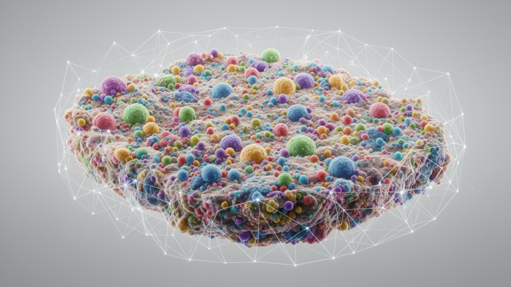
The mathematical pipeline begins by constructing an undirected spatial graph where individual cells function as network nodes and spatial proximities define interconnecting edges. Using single-cell coordinates derived from spatial transcriptomics, the system establishes edges among the ten nearest spatial neighbors based on Euclidean distances between cell centroids, applying inverse-distance edge weighting during graph neural network message passing. Node feature vectors integrate microenvironmental composition, represented as one-hot encoded neighboring cell classifications, alongside cell-intrinsic genomic features captured via the top seven principal components of copy number alteration matrices. For neural classification and continuous regression, SPiCe employs a three-layer graph convolutional network equipped with hidden dimensions of 16 and 32, rectified linear unit activations, dropout regularization, and an explicit Moran’s I spatial regularization penalty that counteracts spatial autocorrelation.
Before analyzing complex human malignancies, the investigators rigorously benchmarked the computational architecture across developmental and simulated benchmarks. The system was first deployed on a MERFISH dataset profiling 280,000 cells across 61 mouse motor cortex tissue slices. Graph neural network models trained to predict masked cell identities achieved high predictive accuracy, with area under the receiver operating characteristic curve values exceeding 0.85 for spatially constrained neuronal subtypes, including layer 2/3, 4/5, and layer 6 intratelencephalic and corticothalamic neurons. Conversely, highly dynamic and functionally plastic populations such as pericytes and oligodendrocyte precursor cells yielded substantially lower predictive accuracy, matching the hypothesis that unconstrained, plastic cell types exhibit reduced predictability from spatial context. Subsequent benchmarking across 100 synthetic spatial transcriptomics datasets confirmed that predefined epithelial and mesenchymal states were consistently recovered alongside their simulated microenvironmental dependencies, whereas hybrid states remained substantially less predictable.
The researchers subsequently deployed SPiCe on a primary human breast cancer specimen profiled with subcellular Xenium in situ spatial transcriptomics, encompassing 167,780 cells and 307 targeted genes, paired with matched whole-transcriptome Visium spatial sequencing. Clonal architecture was resolved by reconstructing subclonal copy number alterations with SCEVAN on the matched Visium data, revealing eight distinct tumor subclones marked by signature chromosomal alterations such as 8q24.3 amplification spanning the MYC oncogene, 17q22-24 amplification, and 11q13.4-q25 deletion. Principal component analysis reduced these alterations into orthogonal axes capturing pivotal driver alterations including CDKN2A, BRCA2, ATM, EGR1, and ERBB2 variations. Epithelial-mesenchymal plasticity was operationalized by scoring a 195-gene hallmark signature from MSigDB, categorized into four balanced quartiles: epithelial, hybrid-epithelial, hybrid-mesenchymal, and mesenchymal states. To prevent misclassification arising from shared transcriptomic features between mesenchymal cancer cells and stromal fibroblasts, the authors used single-cell RNA-sequencing data to identify LUM as a discriminative marker, filtering out 29,000 ambiguous cells to yield 138,780 highly validated cancer cells.
Predictive classification with the graph neural network demonstrated clear divergence in state stability across breast cancer niches. When evaluated under inductive spatial block cross-validation, the mesenchymal tumor state proved to be the most predictable, reaching a mean area under the curve of 0.71 based on intrinsic genomic copy number alterations alone, and climbing to 0.77 when extrinsic tumor microenvironment composition was incorporated. Combining genomic and microenvironmental features yielded no additive performance gain, indicating that while the microenvironment serves as the dominant external driver of epithelial-mesenchymal plasticity, specific somatic copy number profiles become clonally fixed to lock in mesenchymal phenotypes. In sharp contrast, intermediate hybrid states were the least predictable across all configurations, confirming their status as highly dynamic, unconstrained phenotypic intermediates that can rapidly rewire external dependencies.
Interpretation of the neural network parameters using integrated gradients and GNNExplainer uncovered distinct cellular interaction programs across the plasticity continuum. Epithelial tumor cells resided in protected cores primarily interacting with luminal cancer cells, memory B cells, and ACKR1-positive and CXCL12-positive endothelial cells indicative of homeostatic chemokine signaling. Progression toward hybrid and mesenchymal phenotypes coincided with a progressive rewiring of the microenvironment, marked by increased co-option of basal cancer phenotypes, inflammatory cancer-associated fibroblasts, and LYVE1-positive and RGS5-positive endothelial cells associated with invasion and hypoxia. In the fully mesenchymal state, cancer cells formed prominent topological associations with cycling T cells, natural killer T cells, and myofibroblastic cancer-associated fibroblasts, while showing specific statistical links to genomic principal components involving CD44 and ERBB2 alterations.
To overcome the black-box limitations of deep neural networks and measure spatial scale, the team applied spatial error models, geographically weighted regression, and multiscale geographically weighted regression to continuous transition scores. While graph neural networks achieved superior overall classification accuracy, spatial error modeling confirmed that tumor microenvironment composition explained twice the variance in plasticity scores compared to genomic copy number alterations alone. Spatial error modeling across histopathological compartments revealed that invasive tumor areas correlated strongly with advanced mesenchymal progression. Notably, myoepithelial cells exhibited divergent regional behaviors, associating with elevated plasticity in non-invasive ductal carcinoma in situ regions but correlating with reduced plasticity in invasive zones, reflecting their complex transition from structural basement membrane guardians to signaling modulators.
Multiscale geographically weighted regression provided critical insights into the operational spatial ranges over which different stromal and immune populations influence neoplastic plasticity. Unlike standard geographically weighted regression, which enforces a single uniform bandwidth across all predictors, multiscale modeling estimates independent, feature-specific spatial bandwidths representing the effective number of nearest neighbors involved in local interactions. This spatial scale decomposition revealed that myoepithelial cells operate across compact, short-range bandwidths. In striking contrast, myofibroblastic cancer-associated fibroblasts displayed expansive, long-range bandwidths, exerting extended spatial influence across tissue regions through extracellular matrix remodeling, collagen reorganization, and tissue stiffening. Furthermore, multiscale coefficients for CD8-positive T cells precisely mapped their progressive enrichment along the histological continuum from ductal carcinoma in situ toward invasive carcinoma.
The investigators corroborated the generalizability of their framework by analyzing independent five-thousand-gene Xenium datasets profiling an additional human breast tumor and an invasive lung adenocarcinoma. Across both independent malignancies, hybrid states remained consistently the least predictable, whereas mesenchymal phenotypes retained superior predictability with median area under the curve values surpassing 0.80. Mesenchymal tumor cells repeatedly correlated with fibroblasts, endothelial networks, and immune infiltrates, whereas epithelial states aligned with adjacent homotypic epithelial cells and T-cell populations. Direct comparison against the latent-variable spatial decomposition tool SIMVI showed convergent findings, with five out of seven SIMVI-derived spatial archetypes significantly separating epithelial and mesenchymal compartments, while SPiCe uniquely provided explicit predictive quantification and feature-specific spatial bandwidth estimation.
The study authors emphasize important biological and methodological qualifications regarding the interpretation of their findings. Although mathematical predictability under measurable genomic and cellular features correlates strongly with phenotypic constraint, the researchers caution that prediction performance serves as an operational proxy rather than a direct equivalence to cellular plasticity. Lower predictability can stem from unmeasured biological gradients such as interstitial fluid pressure, hypoxia, or nutrient deprivation, as well as technical noise and stochastic gene expression bursts. Furthermore, the four-state quartile discretization represents a pragmatic modeling choice rather than a complete description of the continuous plasticity spectrum. Because the study represents a computational proof of concept based on spatial transcriptomic observations, the authors note that establishing true directional causality among mutations, microenvironmental niches, and phenotypic shifts will require future longitudinal studies and experimental perturbation screens.
Subject of Research: Integrative spatial modeling of cellular plasticity in cancer transcriptomics
Article Title: SPiCe: integrative spatial modelling of cellular plasticity using graph neural networks and geostatistics
Article References: Withnell, E., Celik, C., & Secrier, M. (2026). SPiCe: integrative spatial modelling of cellular plasticity using graph neural networks and geostatistics. Genome Biology. https://doi.org/10.1186/s13059-026-04350-1
Image Credits: AI Generated
DOI: 10.1186/s13059-026-04350-1
Keywords: cellular plasticity, spatial transcriptomics, graph neural networks, geostatistics, breast cancer, epithelial-mesenchymal transition, tumor microenvironment, cancer-associated fibroblasts, spatial regression, multiscale geographically weighted regression, spatial error models, computational oncology, Xenium, copy number alterations, single-cell analysis
News Source: Nathaniel Bowman. (November 19, 2026). Spatial Framework Maps Breast Cancer Cellular Plasticity Using Neural Networks and Geostatistics. Scienmag.