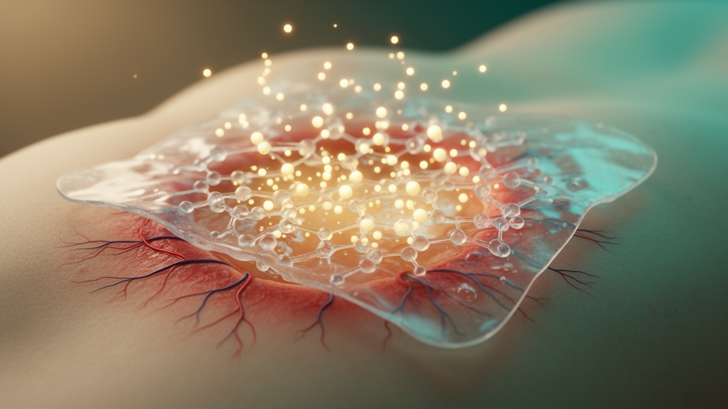
Chronic and severe skin wounds remain one of the most stubborn challenges in regenerative medicine, and a research team in China now reports a deceptively simple material that co-opts the wound’s own hostile chemistry to speed repair. Writing in Frontiers in Pharmacology, scientists at The Affiliated Huaian No. 1 People’s Hospital of Nanjing Medical University describe a reactive oxygen species-responsive hydrogel made from gelatin methacryloyl, or GelMA, loaded with L-arginine. The dressing releases nitric oxide, a short-lived gas molecule with documented roles in blood vessel growth, immune regulation and tissue remodeling, only when it encounters the oxidative burst that characterizes inflamed tissue. In a rat model of full-thickness skin defects, wounds treated with the hydrogel reached a healing rate of 93.7 percent by day 14, substantially outperforming both untreated controls and wounds covered with plain GelMA. The work stands out less for exotic components than for elegant restraint: no nanoparticles, no lasers, no multi-layer assembly, just two biocompatible ingredients and the wound’s own hydrogen peroxide doing the triggering.
Nitric oxide has long been recognized as a molecular multitasker in cutaneous repair. During the inflammatory phase, it governs leukocyte chemotaxis and the secretion of inflammatory mediators; later, it drives vasodilation and stimulates the proliferation, migration and differentiation of endothelial cells that line new blood vessels. In the proliferative phase it enhances fibroblast migration and proliferation, accelerating collagen synthesis and deposition, while also modulating keratinocyte growth and epidermal regeneration. It even carries bactericidal capacity against invading pathogens. The problem has always been delivery. Oral L-arginine supplementation, the amino acid from which nitric oxide synthase enzymes produce the gas, fails to achieve effective local accumulation at the wound site, so systemic doses are diluted across the body before they can meaningfully influence a two-centimeter lesion. Immobilizing the precursor inside a dressing that sits directly on the injury is the logical workaround, and it is precisely what the Huaian team built.
The fabrication is deliberately straightforward. The researchers dissolved GelMA monomer in deionized water with 0.25 percent lithium phenyl-2,4,5-trimethylbenzoylphosphinate, a photoinitiator, then added L-arginine at 0.05 grams per 10 milliliters of pre-crosslinking solution. After sterile filtration through a 0.22-micrometer filter, the mixture was cast into round molds and cured under 405-nanometer visible light at a working distance of three centimeters, yielding uniform disk-shaped dressings two centimeters in diameter. Scanning electron microscopy of freeze-dried cross-sections confirmed that both pure and L-arginine-loaded gels retained a highly porous, interconnected network architecture, the feature that permits nutrient diffusion and cell infiltration in tissue engineering. Fourier-transform infrared spectroscopy indicated that L-arginine binds to the GelMA matrix primarily through physical rather than chemical bonds, which matters because covalent modification of the gelatin backbone would complicate manufacturing and batch consistency.
Equally important, the additive left the hydrogel’s bulk behavior untouched. Swelling experiments in pH 7.4 phosphate-buffered saline at 37 degrees Celsius, conditions simulating wound exudate, showed rapid water uptake in both formulations that plateaued after roughly 20 hours, with no meaningful difference between them. Degradation profiles over 14 days likewise overlapped almost completely, rising slowly at first and accelerating after day 10. That combination of stable swelling and matched degradation is not incidental; it ensures the dressing keeps absorbing exudate while dissolving at roughly the pace of tissue remodeling. For a material intended as a clinical wound dressing, preserving the host scaffold’s properties while grafting on a therapeutic function is the entire engineering challenge, and here the team appears to have passed that test cleanly.
The clever part is the release mechanism. Under oxidative conditions, L-arginine undergoes a reactive-oxygen-mediated oxidation that liberates nitric oxide non-enzymatically. When the researchers exposed the hydrogels to hydrogen peroxide at 50, 100 and 200 micromolar concentrations at 37 degrees Celsius, pure GelMA produced virtually no detectable nitric oxide, confirming that L-arginine is the required substrate. All loaded gels displayed an initial rapid burst of the gas followed by slow, sustained accumulation, and the generation rate scaled positively with peroxide concentration. The chemistry is selective in a biologically meaningful way: among reactive oxygen species found in inflammatory wounds, hydrogen peroxide is the primary trigger for non-enzymatic nitric oxide production from L-arginine, whereas superoxide cannot oxidize the amino acid and instead scavenges newly formed nitric oxide to make peroxynitrite, and hydroxyl radicals non-specifically degrade it without generating the gas at all.
The reaction is also pH-dependent in a way that favors clinical use. Weakly acidic conditions accelerate oxidation of L-arginine’s guanidinium group and promote nitric oxide release, while strongly alkaline environments suppress it. Because the inflammatory wound microenvironment maintains a near-neutral pH, the hydrogel’s trigger chemistry operates in its comfortable range without external assistance. No near-infrared laser irradiation is needed, which the authors note avoids potential thermal damage to peri-wound tissue and eliminates the need for auxiliary equipment at the bedside. In vivo measurements on day 3 after treatment bore the mechanism out: wounds treated with the composite hydrogel showed significantly decreased endogenous hydrogen peroxide and markedly elevated local nitric oxide compared with controls, direct evidence that immobilized L-arginine consumed wound-derived peroxide to generate the gas in situ.
The oxidative stress data reinforce that picture. Three days after treatment, the researchers homogenized wound tissue and quantified reduced glutathione, malondialdehyde and hydrogen peroxide with commercial assay kits. The plain GelMA group showed no significant changes in any of the three markers relative to untreated controls. The composite hydrogel, by contrast, significantly increased glutathione content while sharply reducing malondialdehyde and peroxide levels, all with p-values below 0.001. In other words, the dressing did not merely deliver a signaling molecule; it simultaneously buffered the oxidative damage that peroxide inflicts on regenerating tissue, converting a hostile biochemical environment into a more permissive one for repair.
Functional outcomes followed. In thirty 8-week-old male Sprague-Dawley rats bearing two-centimeter full-thickness dorsal wounds, the composite group consistently showed the smallest residual wound area and highest healing rate from day 3 onward, reaching 93.7 percent closure by day 14, significantly better than both controls and plain GelMA. Histology told a story of quality as well as speed. Hematoxylin and eosin staining revealed a continuous, well-organized epidermis in the treated group, with epidermal thickness of 76.3 plus or minus 18.5 micrometers compared with 119.3 plus or minus 28.0 micrometers in controls, suggesting more physiological re-epithelialization rather than thickened coverage. Masson’s trichrome staining showed dense collagen fibers aligned parallel to the epidermal layer, with a collagen area fraction of 62.6 percent versus 27.0 percent in controls, and the treated wounds showed visible regeneration of sebaceous glands and hair follicles.
Immunofluorescence illuminated the underlying biology. Staining for CD31, an endothelial marker, and alpha-smooth muscle actin showed the highest vessel density in the composite group, reaching 116 vessels per square millimeter, with strong co-localization of the two markers indicating mature vasculature rather than nascent capillaries. Macrophage staining told a complementary story: the ratio of CD206-positive, anti-inflammatory M2 macrophages to total CD68-positive macrophages was significantly elevated in treated wounds, and in vitro experiments with LPS-stimulated RAW 264.7 macrophages confirmed that the L-arginine-loaded gel downregulated the M1 marker CD86 and upregulated CD206, shifting the immune balance toward resolution and repair. Major organ histology at day 14 showed no inflammation, necrosis or pathological abnormalities in heart, liver, spleen, lung or kidney, supporting systemic biosafety in this model.
The authors are candid about limits. The study cannot fully disentangle L-arginine’s nutritional contribution to collagen synthesis from the effects of ROS-triggered nitric oxide, downstream molecular signaling pathways remain unexplored, and long-term in vivo evaluation is absent. The system also lacks anti-biofilm activity, which the team flags as a key optimization target for severely infected ulcers, and efficacy in bacteria-infected or chronic pathological wounds has yet to be systematically verified. Large-animal models and refined spatiotemporal control of nitric oxide release across the distinct phases of healing are the stated next steps. Still, the core proposition is compelling: a one-step, visible-light-crosslinked dressing with no inorganic nanoparticles, no external activation hardware and low production cost, that reads the chemical state of the wound and responds by releasing a molecule that healing tissue can use. If the rat results translate to larger models and eventually patients, the humble amino acid in a gel matrix may become a routine tool for stubborn wounds.
Subject of Research: A ROS-responsive GelMA/L-arginine hydrogel for localized nitric oxide delivery in rat skin wound healing
Article Title: ROS-responsive GelMA/L-Arginine hydrogel with locally sustained NO release for accelerated skin wound healing
Article References: Chen, Y., Wu, D., Wu, H., Yu, G., Yang, W., Wang, Q., Luo, W., & Wang, J. (2026). ROS-responsive GelMA/L-Arginine hydrogel with locally sustained NO release for accelerated skin wound healing. Frontiers in Pharmacology, 17, Article 1970915. https://doi.org/10.3389/fphar.2026.1970915
Image Credits: AI Generated
DOI: 10.3389/fphar.2026.1970915
Keywords: hydrogel, nitric oxide, GelMA, L-arginine, ROS-responsive, wound healing, angiogenesis, macrophage polarization, oxidative stress, biomaterials, rat model, drug delivery
News Source: Bethany Barker. (October 27, 2026). Smart Hydrogel Turns Wound Chemistry Into Healing Nitric Oxide. Scienmag.