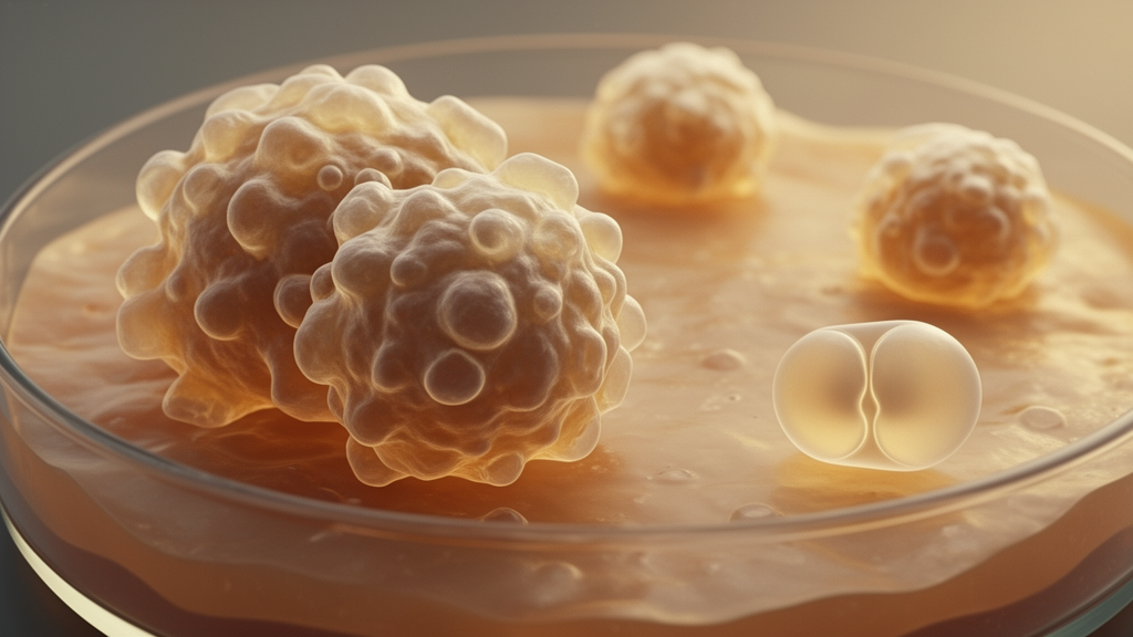
A literature review concludes that the evidence is stronger for cellular senescence as a causal contributor to age-related functional decline than for somatic DNA mutations themselves driving organismal aging. The comparison is based largely on animal experiments, including genetic and pharmacological interventions, rather than large, controlled human trials. Senescent cells can impair tissue regeneration, promote inflammation and disturb neighboring tissues, and removing them has improved several age-related outcomes in mice. Somatic mutations accumulate with age and can affect tissue function, but direct evidence that the mutations are both necessary and sufficient to produce widespread age-related decline remains limited.
The review, published in Aging on August 26, 2026, was written by Lucrezia A. Trastus and Fabrizio d’Adda di Fagagna of the IFOM ETS–The AIRC Institute of Molecular Oncology in Milan, Italy. d’Adda di Fagagna is also affiliated with the Istituto di Genetica Molecolare of the Consiglio Nazionale delle Ricerche in Pavia. Its scope is a comparison of cellular senescence and somatic mutations as possible causes of aging, with necessity and sufficiency used as criteria for causal inference. The authors assess experimental interventions, transplantation studies, cross-species associations, observations in human tissues and inherited conditions, and the biological relationship among DNA damage, damage signaling, senescence and mutation.
Cellular senescence is characterized by stable arrest of cell proliferation. When senescent cells accumulate, they can reduce the capacity of tissues to repair themselves, particularly when stem cells are affected. Their effects may also extend beyond the cells in which they arise. Senescent cells release a complex program of signaling molecules, including inflammatory cytokines, chemokines and tissue-remodeling factors, collectively described as the senescence-associated secretory phenotype, or SASP. These factors can alter surrounding tissues, influence neighboring cell behavior, promote senescence in nearby cells, disrupt extracellular matrix and intercellular barriers, and contribute to fibrosis. The review links these mechanisms to aging hallmarks including telomere dysfunction, chronic inflammation and stem-cell exhaustion.
Experimental findings support a causal role for senescent cells in age-related dysfunction. Removing senescent cells from genetically engineered mice has delayed or reduced several age-related pathologies and improved healthspan; one study also reported increases in median and maximal lifespan. Pharmacological elimination of senescent cells using senolytic approaches has extended these findings in experimental models. The direction of these results is important because the interventions target a candidate cause rather than merely documenting that senescent cells become more common as organisms age. At the same time, the evidence remains predominantly animal-based, and the review notes that sustained clinical benefits of senolytic therapies have not yet been established in large, well-controlled human trials.
Transplantation experiments provide complementary evidence. Relatively small numbers of senescent cells have been able to impair physical function or induce frailty and cognitive decline in young mice. These findings show that introducing senescent cells can contribute to adverse outcomes without requiring the extensive accumulation that accompanies natural aging. They also underscore the importance of the SASP, through which senescent cells can propagate signals through autocrine and paracrine routes. Its inflammatory components can contribute to chronic, low-grade inflammation, while its tissue-remodeling factors can alter the environment around healthy cells. Together, these effects provide a mechanism through which limited numbers of senescent cells could influence stem and immune compartments and broader tissue function.
Evidence connecting somatic DNA mutations to aging is more limited. Mutations accumulate in normal tissues throughout life, and some mutational signatures increase in an approximately clock-like manner with age. Across mammalian species, maximum lifespan is inversely correlated with the rate of somatic mutation accumulation, while DNA-repair capacity has also been associated with longevity. Age-associated mutant clones have also been observed in apparently normal skin, esophagus and blood. Nevertheless, these findings do not by themselves show that altered DNA sequences cause functional decline. The review also notes that some people with inherited defects that substantially increase somatic mutation burden do not display premature-aging phenotypes, which complicates a simple interpretation of mutation accumulation as a universal driver.
Mutations could nevertheless affect aging through several mechanisms. They may disrupt gene regulation and increase transcriptional noise, impair stem and immune cell function, or permit mutant cells to expand clonally. Such expansion creates genetic mosaics within tissues, with age-associated mutant clones detected even in tissues that appear normal. The central difficulty is demonstrating direct causation at the organism level. Experimental evidence that somatic mutations are both necessary and sufficient to produce widespread functional decline remains limited, and available interventions do not readily distinguish the effect of a changed DNA sequence from the damage signaling that accompanied the original lesion.
DNA damage, DNA-damage signaling and DNA mutation are related but distinct events. DNA damage can activate signaling pathways that promote cellular senescence, while failed or inaccurate repair can convert that damage into a permanent mutation. Age-related dysfunction could therefore result from the mutations, from signaling generated by pre-mutation damage, or from both. The review cites experimental models in which inhibiting components of the DNA-damage response has attenuated certain accelerated-aging phenotypes or improved age-related tissue dysfunction without necessarily removing the underlying source of damage. DNA lesions can also interfere directly with replication and transcription, potentially contributing to senescence, stem-cell exhaustion and changes in gene expression. Experiments designed to alter DNA sequence while minimizing accompanying damage signaling would provide a more direct test of whether mutations themselves drive aging.
Major uncertainties remain on both sides of the comparison. Evidence supporting senescence as a causal contributor comes predominantly from animal models, while large, well-controlled human trials have not yet established sustained clinical benefits from senolytic therapies. For somatic mutations, the key unresolved issue is whether altered DNA sequences are primary drivers of organismal aging, rather than markers of damage that also activates senescence and other age-related pathways. The review’s conclusion is therefore comparative rather than absolute: cellular senescence currently has stronger experimental support as a contributor to age-related decline, particularly through its effects on regeneration and its broader effects through the SASP. Somatic mutations clearly accumulate with age and may affect tissue function, but their direct causal role remains unresolved.
Subject of Research: Comparative causal evidence for cellular senescence and somatic DNA mutations in aging
Article Title: Evidence for cellular senescence as a driver of aging stronger than for somatic mutations
Article References: Evidence for cellular senescence as a driver of aging stronger than for somatic mutations. Original publication (Bibliographic details unavailable.)
Image Credits: AI Generated
DOI: Not provided
Keywords: cellular senescence, somatic mutations, aging, DNA damage, senescence-associated secretory phenotype, senolytic therapies, healthspan, genetic mosaics, tissue regeneration, causal inference, Evidence, cellular
News Source: Beatrice Stafford. (October 26, 2026). Review finds stronger causal evidence for senescence than mutations in aging. Scienmag.