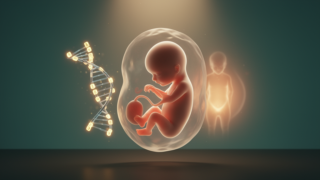
Polycystic ovary syndrome, the most common reproductive and endocrine disorder affecting women of reproductive age, has been renamed polyendocrine metabolic ovarian syndrome (PMOS) in a recent global consensus, a change intended to capture the condition’s endocrine, metabolic and ovarian pathophysiology rather than its ovarian appearance alone. A new review published in Frontiers in Endocrinology by Mahdi, Qannan and El Hajj of Hamad bin Khalifa University in Doha brings together developmental biology, genetics and epigenetics to argue that the syndrome is best understood as a developmental disorder, one whose roots may be set before birth and whose full expression may wait until puberty.
The authors frame their argument around the developmental origins of health and disease (DOHaD) theory, which holds that environmental exposures during critical windows of development can produce lasting changes in tissue structure and function. The fetus, they note, is most vulnerable during the embryonic period, when it uses intrauterine stressors to adjust its phenotype to the environment it predicts it will encounter. This developmental plasticity is mediated at least in part by epigenetic mechanisms, chemical modifications such as DNA methylation and histone changes that modulate gene expression without altering the underlying DNA sequence.
The first formal hypothesis framing the syndrome as the outcome of developmental programming was proposed by Abbott and colleagues in 2002, suggesting that early, possibly prenatal, hyperandrogenism may program the hypothalamic-pituitary-ovarian axis and, together with genetic and metabolic factors, predispose an individual to the condition. The review adopts a “two-hit” model: in-utero developmental programming constitutes the initial priming event, potentially involving epigenetic modifications established during fetal development, while a second hit may be triggered by postnatal environmental factors and the onset of puberty, unmasking vulnerabilities laid down in utero.
Animal models provide much of the strongest evidence for prenatal androgen exposure as a priming factor. In prenatally androgenized rhesus monkeys, exposure produces disordered ovarian function, impaired fertility, hypersecretion of luteinizing hormone and insulin resistance, closely replicating the human syndrome. Exogenous testosterone in rats disrupts early ovarian follicular dynamics and produces the disordered folliculogenesis characteristic of the polycystic ovary. Timing matters: in rhesus monkeys, exposure during early to mid-gestation may produce more pronounced ovarian and PMOS-like phenotypes than later exposure. In humans, however, the evidence is less direct. Maternal hyperandrogenemia may not be sufficient to program the syndrome in female offspring, because the human fetus is partially protected through placental aromatization of androgens to estrogens and high concentrations of sex hormone-binding globulin during pregnancy. Observational studies have suggested an association between the condition and elongated anogenital distance, a possible clinical marker of prenatal androgen exposure, but biochemical studies of cord blood androgens in daughters of affected women have reported higher, lower or unchanged concentrations, and cord blood obtained at delivery may not reflect androgen exposure during the critical early-to-mid-gestation window.
An alternative or complementary pathway may run through maternal metabolic disturbances. Maternal obesity, insulin resistance and hyperglycemia may alter the placental metabolic and endocrine environment and, in genetically susceptible female fetuses, contribute to fetal hyperinsulinemia, which in turn may promote ovarian androgen production and disrupt folliculogenesis in utero. The review also highlights endocrine-disrupting chemicals such as bisphenol A, for which epidemiological studies have reported higher body fluid concentrations in women with PMOS compared with controls, and experimental rodent studies suggest effects on ovarian steroidogenesis, hypothalamic-pituitary-gonadal axis function and epigenetic regulation. The authors caution, however, that the causal direction of the human cross-sectional association remains unresolved: metabolic disturbances in patients, such as increased adiposity, may promote the accumulation of these chemicals, meaning higher measured concentrations could reflect altered distribution and elimination rather than higher external exposure.
Genetics alone cannot explain the syndrome’s heterogeneity. Familial studies reported a 20 to 40 percent prevalence among first-degree relatives compared with background prevalence, and early twin studies estimated that genetic factors accounted for up to 79 percent of variation, although the authors note these estimates were likely inflated because monozygotic twins also share more similar intrauterine environments and postnatal lifestyles. Genome-wide association studies have identified risk loci, with the first two studies in China identifying 11 loci, two of which were replicated in European-ancestry patients, and the most recent meta-analysis identifying 29 independent loci across ancestries. Yet identified loci explain only up to 10 percent of PMOS heritability, and GWAS identifies loci rather than genes, creating biological uncertainty. This gap is precisely where the authors place epigenetics.
The review devotes particular attention to the neuroendocrine machinery that puberty activates. Pulsatile release of gonadotropin-releasing hormone from the hypothalamus drives pituitary secretion of luteinizing hormone and follicle-stimulating hormone, and the kisspeptin-neurokinin B-dynorphin (KNDy) neuron system in the arcuate nucleus plays a central role in regulating this pulsatility. Epigenetic regulation of the KISS1 gene, which encodes kisspeptin, is described as a key player in the initiation of puberty: the shift of KISS1 from a repressed to an active state in hypothalamic neurons enables GnRH pulsatility, a switch mediated by coordinated removal of Polycomb repressive complexes and activation of Trithorax group proteins. In women with PMOS, feedback of estradiol and progesterone on LH pulsatility is much weaker than in healthy women, in whom these hormones dampen LH pulsatility by roughly 60 percent, and blocking androgen receptors with flutamide has been shown to restore normal feedback sensitivity. The authors are careful to note that there is currently no direct evidence that prenatal exposures induce specific KISS1 chromatin alterations in the human hypothalamus; the mechanistic framework rests largely on experimental animal studies, and direct characterization of KISS1 chromatin states in human hypothalamic tissue remains inherently challenging.
Metabolic dysfunction features prominently in the proposed framework. About 70 percent of women with PMOS show clinically measurable insulin resistance independent of body mass index, characterized by impaired metabolic insulin signaling in skeletal muscle and adipose tissue while the ovaries remain relatively sensitive to insulin, stimulating theca-cell androgen production. The review connects this to the gut microbiome through the dysbiosis theory proposed by Tremellen and colleagues, in which a poor-quality diet weakens intestinal barrier integrity, allowing bacterial lipopolysaccharide to enter the circulation and trigger chronic low-grade inflammation and impaired insulin signaling. Animal studies support the pathway: in a letrozole-induced rat model, LPS administration increased ovarian inflammatory markers and impaired reproductive function, and a DHEA-induced mouse model showed compromised intestinal barrier integrity and upregulated ovarian inflammatory signaling that were reversed with dietary modification. Human observational findings are mixed. One study of 73 women with PMOS and 48 controls found significantly lower gut microbial alpha diversity in patients, while a recent systematic analysis found no significant alpha-difference but distinct microbial composition varying with testosterone levels. The authors stress that the causal direction of the gut-inflammation-insulin resistance axis in humans remains uncertain.
The framework also engages with the 2023 International Evidence-based Guideline for PCOS. Anti-Müllerian hormone, secreted by granulosa cells of small antral follicles, is now used as an alternative to ultrasound-based polycystic ovarian morphology in adult diagnostic criteria, with meta-analyses showing pooled sensitivity of 0.79 and specificity of 0.87 in adults. In adolescents, however, AMH performs considerably worse, with pooled sensitivity and specificity of 0.66 and 0.78, and is not endorsed for diagnosis. Longitudinal studies show the predictive value of elevated AMH or polycystic ovarian morphology in adolescent girls is less than 22 percent, whereas adolescents with oligomenorrhea are approximately 4.4 times more likely to develop PMOS in adulthood. The guideline therefore recommends postponing definitive diagnosis until around eight years after menarche, aligning with full maturation of the hypothalamic-pituitary-ovarian axis, and identifying girls with suggestive features as “at risk” for later reassessment. The guideline also endorses healthy lifestyle behaviors for all women with PMOS, and the review notes animal evidence that treadmill exercise and alternate-day feeding downregulate DNA methyltransferase expression, improve glucose tolerance and ovarian function, and modulate histone acetylation, suggesting lifestyle interventions might reshape epigenetic regulation.
The authors acknowledge that the field’s overwhelming reliance on cross-sectional designs cannot determine causal direction, and that the most comprehensive systematic review of longitudinal research identified only 21 longitudinal studies encompassing 62,123 participants, with follow-up ranging from 4 weeks to 32 years, concluding that existing longitudinal data are inadequate and conflicting. What the field urgently requires, they argue, is a prospective longitudinal birth cohort study, ideally in daughters of women with PMOS, with epigenomic profiling of cord blood and peripheral blood during puberty, alongside prenatal exposures, hormonal profiles, AMH trajectories and dietary patterns. Such a design could determine the relative contributions, timing and reversibility of prenatal versus peripubertal epigenetic programming, and whether puberty primarily unmasks or actively promotes the epigenetic alterations that give the syndrome its final form.
Subject of Research: A review of the developmental, genetic and epigenetic mechanisms proposed to underlie polyendocrine metabolic ovarian syndrome
Article Title: The developmental, genetic, and epigenetic basis of polyendocrine metabolic ovarian syndrome
Article References: Mahdi, A., Qannan, A., & El Hajj, N. (2026). The developmental, genetic, and epigenetic basis of polyendocrine metabolic ovarian syndrome. Frontiers in Endocrinology, 17, Article 1924187. https://doi.org/10.3389/fendo.2026.1924187
Image Credits: AI Generated
DOI: 10.3389/fendo.2026.1924187
Keywords: PCOS, PMOS, epigenetics, DOHaD, AMH, gut microbiome, insulin resistance, puberty, GWAS, hyperandrogenism, KNDy neurons, developmental programming
News Source: Scarlett Davidson. (October 28, 2026). PCOS reframed as PMOS: fetal programming, epigenetics and puberty’s second hit. Scienmag.